
Method and device for automatically splicing image sequences and splicing system

A splicing device and splicing position technology, which is applied in image enhancement, image analysis, image data processing, etc., can solve problems such as distortion of shooting conditions, time-consuming search, and difficulty in correct splicing of images, and achieve the effect of simple and effective splicing

Active Publication Date: 2010-06-16
SHENZHEN MINDRAY BIO MEDICAL ELECTRONICS CO LTD
View PDF0 Cites 28 Cited by
  • Summary
  • Abstract
  • Description
  • Claims
  • Application Information

AI Technical Summary

Problems solved by technology

However, due to the limited size of imaging devices such as flat panel detectors (for DR systems) or imaging plates (for CR systems) in X-ray imaging systems, as well as other limitations of existing imaging techniques, only large-sized objects Take multiple body position shots, and then use image processing technology to synthesize the obtained body position images into a panoramic image
[0003] However, multiple shootings will cause the following problems: First, the exposure doses of two exposures in different body positions are different, resulting in inconsistent gray distributions in overlapping areas of adjacent images; Changes in voluntary movement, peristalsis of the large and small intestine, physiological contraction of the bladder, etc.) lead to differences in tissue images taken successively; third, changes in shooting conditions and other factors will cause slight distortion
These problems make it difficult to stitch images correctly
In addition, due to the large amount of data in medical images, the traditional grayscale or gradient global search is very time-consuming, which brings challenges to the dynamic and real-time application of medical images.
[0004] Taking X-ray images as an example, there are two main types of existing stitching technologies, one is to rely on special equipment to set obvious marks on each X-ray image, and then manually or automatically recognize the marks to achieve image stitching; the other The category is automatic stitching, one of which is dependent on the inherent characteristics of the device (such as the overlapping features of CR imaging plates), using different anatomical body images at multiple scales and using different algorithms or parameters to achieve stitching; the other is Image stitching technology that only relies on image features, but the accuracy is not high, often requires manual further correction and correction
In recent years, image panoramic stitching technology has developed rapidly. Although a technique using scale invariant feature transformation theory (Scale Invariant FeatureTransform: SIFT) for panoramic stitching has been proposed, it has not been successfully used in panoramic stitching of X-ray images. No report

Method used


Image

  • Method and device for automatically splicing image sequences and splicing system
  • Method and device for automatically splicing image sequences and splicing system
  • Method and device for automatically splicing image sequences and splicing system

Examples


Embodiment Construction

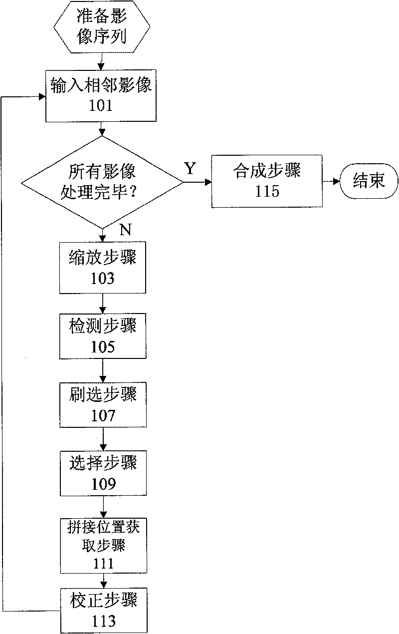
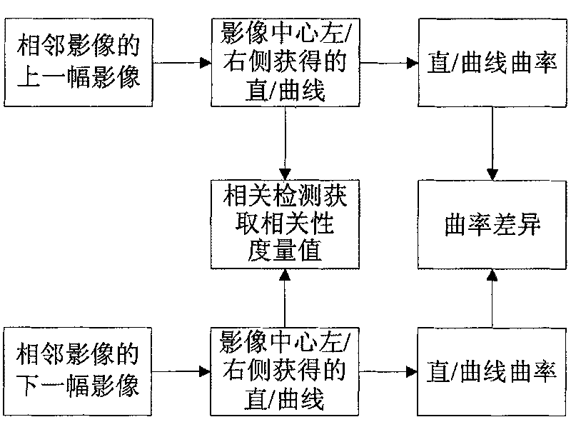
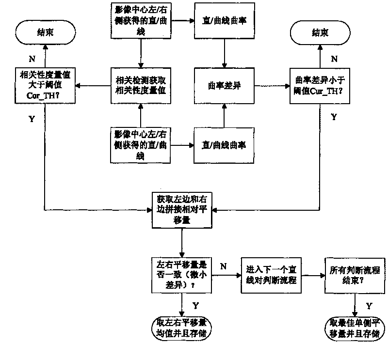
[0019] Such as figure 1 As shown, the image stitching method according to this embodiment mainly includes a detection step 105 , a selection step 109 , and a stitching position acquisition step 111 . In addition, a scaling step 103 , a brushing step 107 , a correcting step 113 , and a compositing step 115 are also optionally included. The specific process of image processing by using the image stitching method according to this embodiment is as follows:

[0020] First, input the adjacent image (step 101), and the image can be scaled to 1 / 2, 1 / 4 or 1 / 8 of the original image according to the needs (step 103), to speed up the calculation speed, and it is generally preferable to use 1 / 8 ; In addition, for images with different magnifications, zoom in and out to make them the same size. If the image pixels are high-bit, such as 12bit or 14bit images, it can be linearly converted to 8bit images. The adjacent images are detected sequentially (step 105), and the splicing displaceme...


PUM


Abstract

The invention discloses a method and a device for automatically splicing image sequences and a splicing system. The method for automatically splicing the image sequence mainly comprises: a step of detection used for detecting organization edges of adjacent images; a step of selection used for computing the similarity of the organization edge lines of the detected adjacent images, and selecting the edge line pair meeting a similarity threshold as an optimized edge line pair; and a step of acquiring splicing positions, namely determining the splicing displacement of the adjacent images in a horizontal direction and in a vertical direction according to the selected optimized edge line pair to determine the splicing positions in the horizontal direction and the vertical direction, wherein the splicing of the adjacent images is realized according to the acquired splicing positions. The method and the device are simply implemented and can effectively splice medical image sequences.

Description

technical field [0001] The present invention relates to an image processing technology, in particular to a method and device for automatically splicing image sequences to synthesize a panoramic image. Background technique [0002] Complete images of large-scale objects (such as X-ray images, or even complete X-ray images of human bodies) play an important role in orthopedic surgery, orthopedic or plastic treatment, etc. However, due to the limited size of imaging devices such as flat panel detectors (for DR systems) or imaging plates (for CR systems) in X-ray imaging systems, as well as other limitations of existing imaging techniques, only large-sized objects Take multiple body position shots, and then use image processing technology to synthesize the obtained body position images into a panoramic image. [0003] However, multiple shootings will cause the following problems: First, the exposure doses of two exposures in different body positions are different, resulting in ...

Claims


Application Information

Patent Timeline
IPC IPC(8): G06T5/50G06T7/00
Inventor 饶玉明
Owner SHENZHEN MINDRAY BIO MEDICAL ELECTRONICS CO LTD
Learn More