Supercharge Your Innovation With Domain-Expert AI Agents!

Early tumor detection method

A detection method and tumor technology, applied in diagnostic recording/measurement, medical science, sensors, etc., to achieve valuable treatment time, improve resolution, and reduce the amount of calculation

Inactive Publication Date: 2010-07-21
NAT UNIV OF DEFENSE TECH
View PDF0 Cites 7 Cited by
  • Summary
  • Abstract
  • Description
  • Claims
  • Application Information

AI Technical Summary

Problems solved by technology

[0004] In order to overcome the deficiencies of the existing tumor detection methods, the present invention provides an early tumor detection method, which can combine the tissue structure partition information of the region of interest obtained by the existing medical detection equipment, and refer to the electromagnetic characteristic parameter distribution database of various tissues of the human body. Different organizational structure partitions assign the value range of electromagnetic characteristic parameters

Method used


Image

  • Early tumor detection method
  • Early tumor detection method
  • Early tumor detection method

Examples


Embodiment Construction

[0019] Embodiments of the present invention will be described in detail below in conjunction with the accompanying drawings.

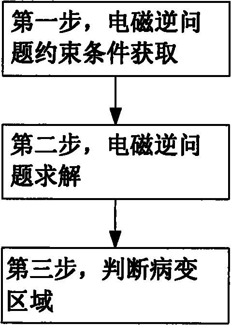
[0020] figure 1 Shown is a schematic flow chart of the early tumor detection method according to the present invention, including three steps of acquiring electromagnetic inverse problem constraint conditions, solving electromagnetic inverse problem and judging lesion area. The first step, the acquisition of the constraints of the electromagnetic inverse problem, includes the acquisition of the boundary value of the electrical signal and the acquisition of organizational structure partition information. The second step, solving the electromagnetic inverse problem, is to use the improved Monte Carlo inversion method to inversely deduce the distribution of tissue electromagnetic characteristic parameters in the region of interest. The third step, judging the lesion area, is to use the existing medical knowledge to manually search for the abnormal area o...


PUM


Abstract

The invention provides an early tumor detection method. The method combines traditional medical detection equipment to obtain tissue partition information of an interest region, distributes the range of electromagnetic property parameters to different tissue structure partitions by referring to the electromagnetic property parameter distribution database of all tissues of the human body, and then works out the electromagnetic property parameter distribution database of the tissue of the interest region through electromagnetic inverse problem solution, and effectively detect early tumors and the tissue lesions without clear boundary. The method combines the tissue partition information with the electromagnetic property parameter distribution range information of the tissue during solving electromagnetic inverse problem, thereby greatly reducing the computation. The electromagnetic inverse problem solving method of the invention which combines with the traditional medical detection equipment can effectively detect the early tumors and the tissue lesions without clear boundary.

Description

technical field [0001] The invention relates to a medical detection method for detecting the existence of tumors, in particular to a medical detection method capable of detecting early tumors and pathological changes of non-obvious border tissues. Background technique [0002] At present, the main tumor detection method is to use existing equipment such as CT, nuclear magnetic resonance, and B-ultrasound for detection. These medical detection devices emit a certain form of energy waves to the human body, then receive signals transmitted or reflected from the human body, and finally generate images through imaging algorithms. Although medical imaging technology has made considerable progress and development, there are still many problems in clinical use, mainly including: ① It is difficult to detect early tumors and tissue lesions without obvious boundaries; The accuracy rate of continuous area detection is not high. [0003] In addition to the traditional tumor detection m...

Claims


Application Information

Patent Timeline
Patent Type & Authority Applications(China)
IPC IPC(8): A61B5/05
Inventor 刘培国丁亮刘继斌周东明李高升
Owner NAT UNIV OF DEFENSE TECH
Features
  • R&D
  • Intellectual Property
  • Life Sciences
  • Materials
  • Tech Scout
  • Unparalleled Data Quality
  • Higher Quality Content
  • 60% Fewer Hallucinations
Learn More