Method for visualizing three-dimensional anatomical tissue structure model of human heart based on ray cast volume rendering algorithm

A tissue structure and volume rendering technology, applied in computing, 3D modeling, image data processing, etc., can solve problems such as the inability to realize the visualization of cardiac anatomy and tissue structure models

Inactive Publication Date: 2010-08-04
HARBIN INST OF TECH
View PDF3 Cites 29 Cited by
  • Summary
  • Abstract
  • Description
  • Claims
  • Application Information

AI Technical Summary

Problems solved by technology

[0003] In order to solve the problem that the visualization of the cardiac anatomical structure model cannot be realized in the prior art, the present invention provides a visualization method for the three-dimensional anatomical tissue structure model of the human heart based on the ray-casting volume rendering algorithm

Method used


Image

  • Method for visualizing three-dimensional anatomical tissue structure model of human heart based on ray cast volume rendering algorithm
  • Method for visualizing three-dimensional anatomical tissue structure model of human heart based on ray cast volume rendering algorithm
  • Method for visualizing three-dimensional anatomical tissue structure model of human heart based on ray cast volume rendering algorithm

Examples


specific Embodiment approach 1

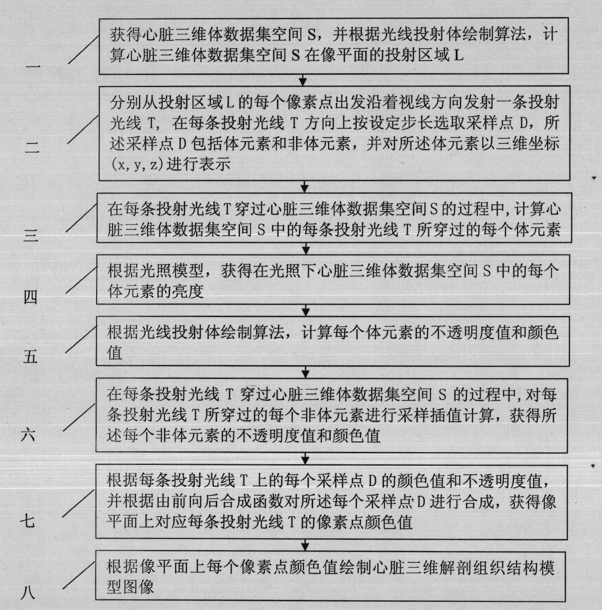
[0015] Specific implementation mode one: according to the instructions attached figure 1 Specifically illustrate this embodiment, the visualization method of the three-dimensional anatomical structure model of the human heart based on the ray-casting volume rendering algorithm described in this embodiment, its specific process is:

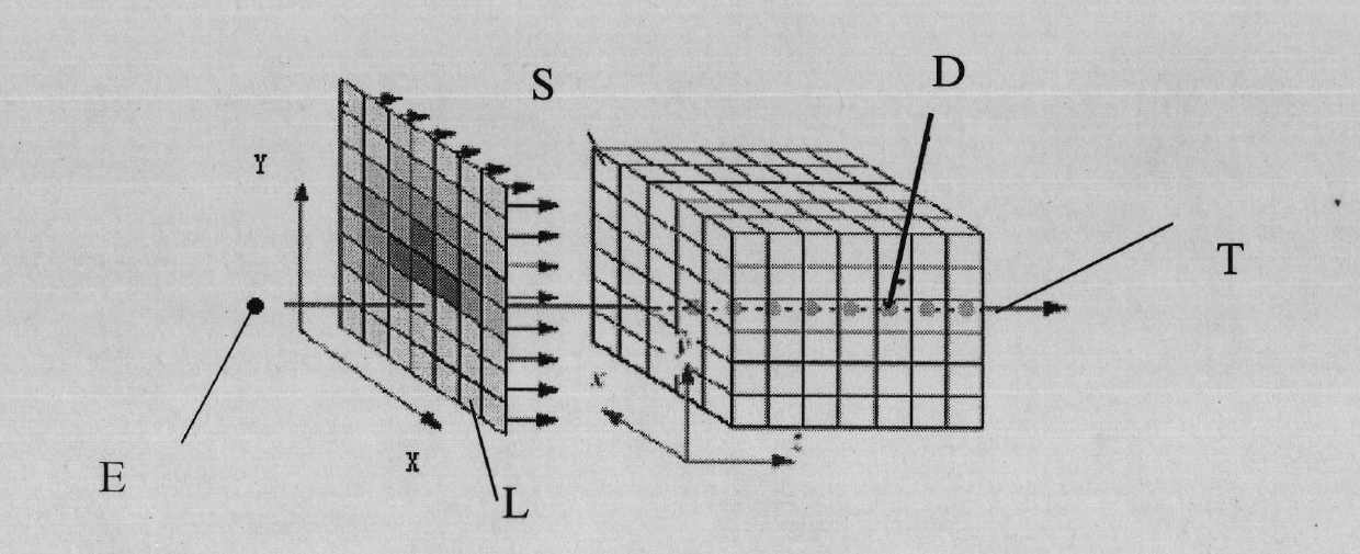
[0016] Step 1: Obtain the heart 3D volume data set space S, and calculate the projection area L of the heart 3D volume data set space S on the image plane according to the ray-casting volume rendering algorithm;

[0017] Step 2: Starting from each pixel point of the projection area L, emit a projection ray T along the line of sight direction, and select a sampling point D in the direction of each projection ray T according to a set step size, and the sampling point D includes voxels and non-volume elements, and represent the volume elements with three-dimensional coordinates (x, y, z);

[0018] Step 3: During the process of each projected ray T pa...

specific Embodiment approach 2

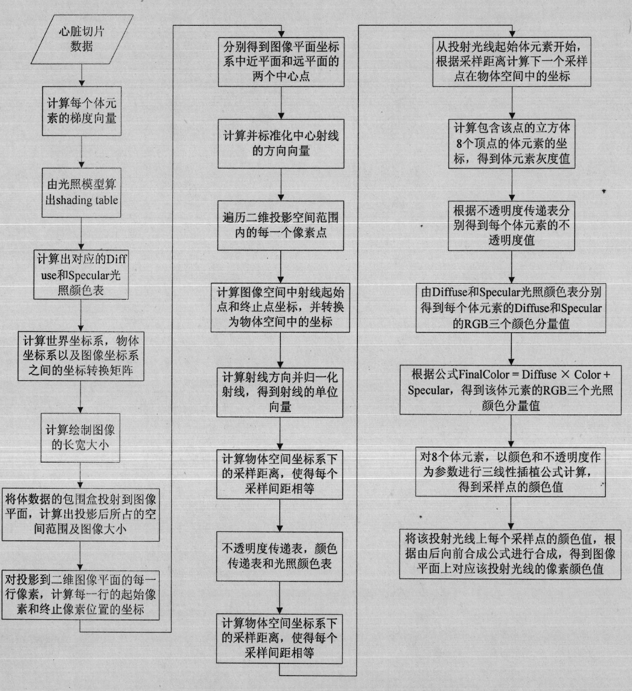
[0024] Embodiment 2: This embodiment is a further description of Embodiment 1. In Embodiment 1, in Step 5, according to the ray-casting volume rendering algorithm, the method for calculating the opacity value and color value of each voxel is:

[0025] Step 511: Using the brightness attribute and gradient attribute of each voxel in the heart three-dimensional volume data set space S as parameters, construct an opacity transfer function, and obtain the opacity of each voxel in the heart three-dimensional volume data set space S Value, the gradient attribute includes gradient value and gradient direction;

[0026] Step 512: Construct a color transfer function, and obtain the color value of the voxel according to the gray intensity of the voxel in the heart tribody dataset space.

specific Embodiment approach 3

[0027] Embodiment 3: This embodiment is a further description of Embodiment 2. In Embodiment 2, in step 511, the brightness attribute and gradient attribute of each voxel in the heart three-dimensional volume data set space S are used as Parameters, to construct the opacity transfer function, the method to obtain the opacity value of each voxel in the heart three-dimensional volume data set space S is: construct the opacity transfer function

[0028]

[0029] Among them, f v Indicates the light intensity of voxels greater than or equal to the corresponding tissue gradient threshold in each tissue in the heart three-dimensional volume data set space S, the tissue includes ventricles and atria, if the gradient value is normalized to [0, 1], then The gradient threshold is 0.8, r represents the ith individual element and the light intensity is f v The maximum distance of voxel elements of , Indicates that the light intensity is I i The gradient value of voxel i, α i (r, f ...


PUM


Abstract

The invention discloses a method for visualizing a three-dimensional anatomical tissue structure model of a human heart based on a ray cast volume rendering algorithm which, relates to the method for visualizing the three-dimensional anatomical model of the human heart and solves the problem of incapability of visualizing the anatomical tissue structure model of the heart in the prior art. The method comprises the following steps of: (1) obtaining a heart three-dimensional volume data set; (2) selecting a sampling point; (3) calculating a gradient of each volume element in a heart three-dimensional volume data set space; (4) obtaining brightness of each volume element in the heart three-dimensional volume data set space under light; (5) calculating an opacity value and a color value of each volume element; (6) obtaining the opacity value and the color value of each volume element; (7) obtaining pixel point color values corresponding to projection light beams on an image plane; and (8) rendering an image of the heart three-dimensional anatomical tissue structure model according to each pixel point color value on the image plane. The invention lays the foundation for exactly simulating the structure appearance and the behaving function of the heart.

Description

technical field [0001] The invention relates to a visualization method for a three-dimensional anatomical model of the human heart, in particular to a visualization method for a three-dimensional anatomical tissue structure model of the human heart based on a light projection volume rendering algorithm. Background technique [0002] The heart is a complex and comprehensive system integrating electrophysiology, hemodynamics, nerve and biological control. Virtual heart models based on a single perspective (anatomical / electrophysiological / mechanical) often cannot effectively reflect the complex interactions between different functions of the heart at a macro level. Therefore, it is difficult to make precise predictions about the behavior and properties of the heart under normal and pathological conditions. Therefore, it is necessary to establish a model of cardiac anatomical tissue structure, so as to integrate it with electrophysiological and mechanical models, so as to achiev...

Claims


Application Information

Patent Timeline
Patent Type & Authority Applications(China)
IPC IPC(8): G06T17/00
Inventor 王宽全杨飞左旺孟袁永峰张宏志
Owner HARBIN INST OF TECH
Why Eureka
Try Eureka