Semi-on-body biomechanical experimental method using cervical structure to simulate extensor muscles behind the neck

A mechanical experiment and biomechanical technology, applied in the field of cervical vertebra biomechanics experiments, can solve the problems of lack of biological changes, errors in experimental results, and the authenticity of model construction.

Active Publication Date: 2010-10-06
YUEYANG INTEGRATED TRADITIONAL CHINESE & WESTERN MEDICINE HOSPITAL SHANGHAI UNIV OF CHINESE TRADITIONAL MEDICINE
View PDF2 Cites 10 Cited by
  • Summary
  • Abstract
  • Description
  • Claims
  • Application Information

AI Technical Summary

Problems solved by technology

However, it is often difficult to obtain physical properties of various soft tissues such as ligaments and intervertebral discs, and even if such data are available, they may not be sufficient for model building
The accuracy of the test results of the 3D finite element model depends entirely on the authenticity of its model construction. Therefore, the imperfect model establishment will inevitably lead to serious errors in the

Method used


Image

  • Semi-on-body biomechanical experimental method using cervical structure to simulate extensor muscles behind the neck
  • Semi-on-body biomechanical experimental method using cervical structure to simulate extensor muscles behind the neck
  • Semi-on-body biomechanical experimental method using cervical structure to simulate extensor muscles behind the neck

Examples


Embodiment Construction

[0038] Experimental method of the present invention comprises the following steps:

[0039] 1. Preparation of in vitro biomechanical models

[0040] (1). Source and production of specimens

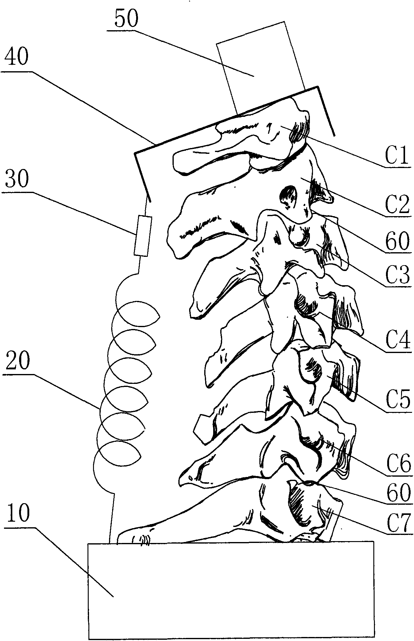
[0041] A human cervical spine that died of acute traumatic brain injury was used. A segment of the spine from the base of the skull to the second thoracic vertebra is amputated within 2 hours of death, such as figure 1 As shown, it includes the first to seventh cervical vertebrae C1-C7 and the first and second thoracic vertebrae. Muscles are removed to avoid damage to ligaments and small joints. The above-mentioned human cervical spine specimens were placed in a natural position, avoiding hyperextension, hyperflexion and rotation, and stored in a low-temperature refrigerator at -40°C. The cervical spine is preserved by this method, and its biomechanical properties will not change (see "Spine" magazine, 1991, No. 16, page 117). Each specimen was taken out from the low-temperature refri...


PUM


Abstract

The invention discloses a semi-on-body biomechanical experimental method using a cervical structure to simulate extensor muscles behind the neck. The method comprises the following steps: cutting out the first to seventh cervical vertebra, the first dorsal vertebra and the second dorsal vertebra from the body of a person who died within 2 hours, removing muscles while avoiding damaging ligaments and facet joints; fixing an upper bearing tray on the first cervical vertebra, fixing a lower bearing tray under the second dorsal vertebra, installing a loading weight on the upper bearing tray; opening 2-3mm of incisions separately at the front junctions and back joints of the third/fourth dorsal vertebra, the fourth/fifth dorsal vertebra, the fifth/sixth dorsal vertebra and the sixth/seventh dorsal vertebra, inserting miniature pressure sensors respectively, sewing with silk threads; connecting a tension sensor and a tension spring between the upper and lower bearing trays to prepare a cervical vertebra biomechanical model; and fixing a pulling-extending force sensor on a cervical vertebra experimental loading table, connecting a wedge-shaped metal block with the upper bearing tray through screws, using the loading table to apply a pulling-extending force and a loading force on the cervical vertebra biomechanical model, and recording the measured values.

Description

technical field [0001] The invention relates to the technical field of biomechanics, in particular to a cervical spine biomechanical experimental method capable of simulating the cervical posterior extensor muscles. Background technique [0002] Cervical spondylosis (Cervical Syndrome, CS) is a common clinical disease. In recent years, it has shown a high incidence and younger trend, and has gradually become a new type of occupational disease. Due to changes in people's work and lifestyle, the probability of neck flexion has greatly increased, causing the soft tissue of the neck and the main structure of the cervical spine to be in a state of fatigue for a long time due to reasons such as working posture and living habits. Under the influence of bad posture, mental stress and other factors, due to the resistance to the preload caused by gravity, the contraction is eccentric, which can cause muscle fiber damage and weaken muscle strength, which directly leads to the destructi...

Claims


Application Information

Patent Timeline
IPC IPC(8): G09B23/28
Inventor 房敏程英武方磊朱清广
Owner YUEYANG INTEGRATED TRADITIONAL CHINESE & WESTERN MEDICINE HOSPITAL SHANGHAI UNIV OF CHINESE TRADITIONAL MEDICINE
Why Eureka
Try Eureka