Modeling method for micropore structure in bionic bone scaffold

A modeling method and technology of microscopic pores, applied in special data processing applications, instruments, electrical digital data processing, etc., can solve problems such as uneven distribution, increased distortion of internal connected pores, and single shape of internal microscopic pores

Inactive Publication Date: 2011-04-20
上海蓝衍生物科技有限公司
View PDF3 Cites 11 Cited by
  • Summary
  • Abstract
  • Description
  • Claims
  • Application Information

AI Technical Summary

Problems solved by technology

In the case of ensuring that the bionic bone scaffold has a high porosity and surface area, improve the single shape and uneven distribution of the internal microscopic por

Method used


Image

  • Modeling method for micropore structure in bionic bone scaffold
  • Modeling method for micropore structure in bionic bone scaffold
  • Modeling method for micropore structure in bionic bone scaffold

Examples


Embodiment Construction

[0053] A preferred embodiment of the present invention is described in detail as follows in conjunction with accompanying drawing:

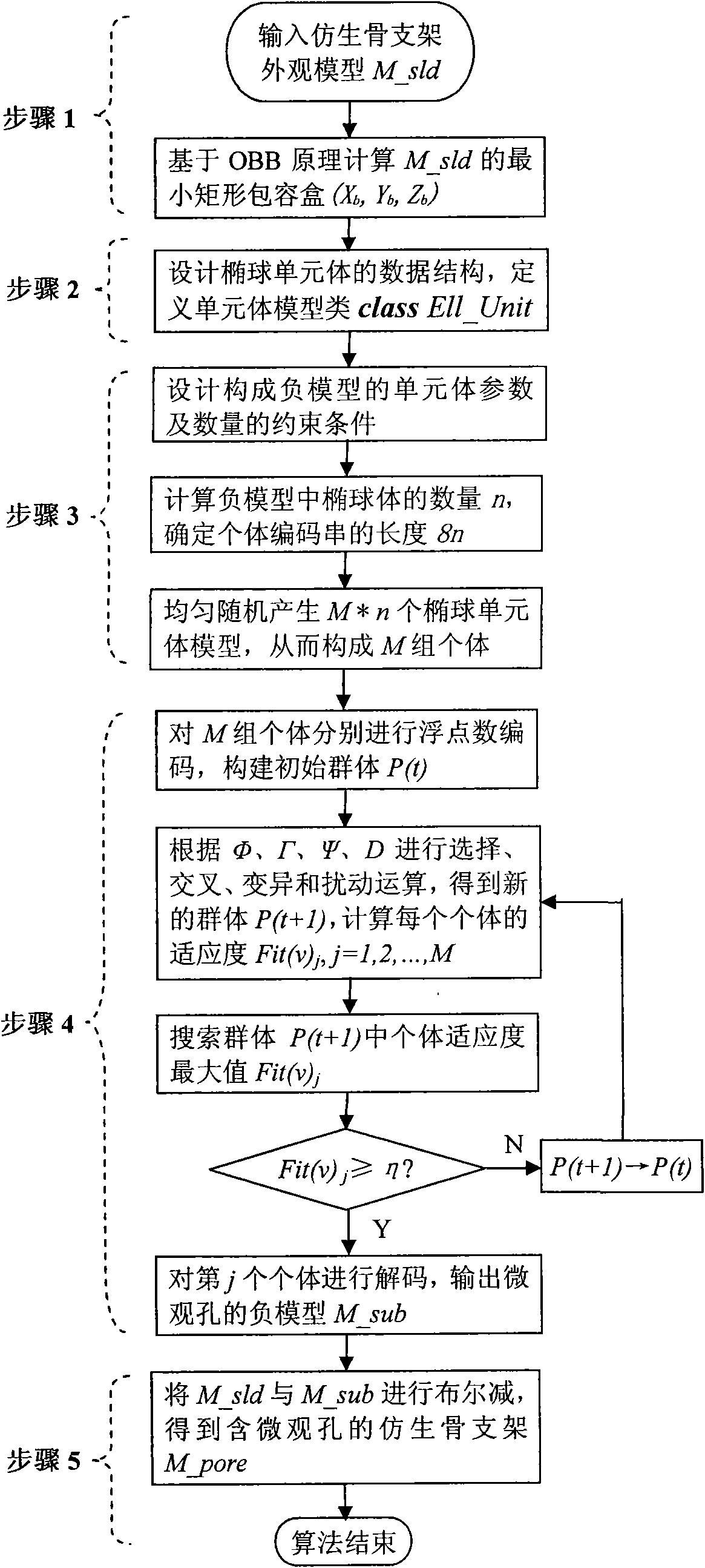
[0054] The specific implementation process of the microscopic pore structure modeling method inside the bionic bone scaffold is as follows: figure 2 As shown, it mainly includes the following five steps:

[0055] Step 1 Calculate the minimum containing box of the bone scaffold model

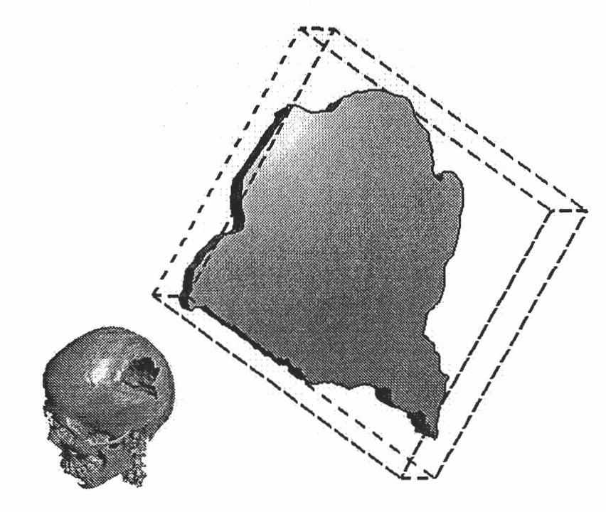
[0056] Input the bionic bone scaffold appearance model M_sld, and calculate the minimum rectangular containing box of M_sld based on the OBB principle (X b , Y b ,Z b ), as the spatial constraint boundary of the microscopic pore structure negative model M_sub;

[0057] Step 2 Construct the unit body model of the microscopic pore structure of the scaffold

[0058] Design the data structure of the ellipsoid unit body according to the model parameter group described in formula (1), and define the unit body model class:

[0059] ELL=(a,β,x c ,y c ,z c , θ x ,...


PUM


Abstract

The invention relates to a modeling method for a micropore structure in a bionic bone scaffold. The method comprises the following steps that: a negative model of the micropore structure in the bionic bone scaffold is solved on the basis of a multi-constraint knapsack problem model by using an ellipsoid as a base element, using the minimum accommodating box of a bone scaffold model as a constraint space and using a hybrid genetic algorithm; and then a bionic bone scaffold model comprising the micropore structure is built through a Boolean subtraction operation between the scaffold model whichdoes not contain micropores and the negative model of the micropore structure. Under the condition of ensuring relatively high void ratio and superficial area of the micropore structure in the bionicbone scaffold, by the method, single shape of the internal micropores and uneven distribution of the micropores are overcome and the torsion resistance of internal communication pore canals in the bionic bone scaffold is increased, so that the performance of the bionic bone scaffold model is improved and the bionic bone scaffold model has relatively high bioactivity.

Description

technical field [0001] The invention relates to a modeling method of a bionic bone support in the field of bone tissue engineering, in particular to a modeling method of a microscopic hole structure inside a bionic bone support. Background technique [0002] In the field of bone tissue engineering research, the co-cultivation of osteoblasts and bionic bone scaffold complexes in vitro is the beginning and key of constructing tissue engineered bones, and it is also a hot research topic at home and abroad. From the perspective of bone tissue engineering, the biomimetic bone scaffold plays the role of bone extracellular matrix, which is a biomimicry of the structure and function of bone extracellular matrix. The biomimetic bone scaffold is a three-dimensional scaffold structure connected by heterogeneous micropores, which creates a microenvironment conducive to the adhesion, growth, proliferation and function of bone cells. Medical research shows that in order to make the bioni...

Claims


Application Information

Patent Timeline
IPC IPC(8): G06F17/50
Inventor 尤飞胡庆夕姚远温婷
Owner 上海蓝衍生物科技有限公司
Why Eureka
Try Eureka