Method for predicting tumor metastasis and invasion capacity in vitro and nucleotide fragments

A transfer ability, nucleotide technology, applied in biochemical equipment and methods, DNA/RNA fragments, microbial determination/inspection, etc.

Inactive Publication Date: 2011-08-03
BEIJING CANCER HOSPITAL PEKING UNIV CANCER HOSPITAL
View PDF0 Cites 5 Cited by
  • Summary
  • Abstract
  • Description
  • Claims
  • Application Information

AI Technical Summary

Problems solved by technology

There is no report on the use of SRF methylation inactivation to measure t

Method used


Image

  • Method for predicting tumor metastasis and invasion capacity in vitro and nucleotide fragments
  • Method for predicting tumor metastasis and invasion capacity in vitro and nucleotide fragments
  • Method for predicting tumor metastasis and invasion capacity in vitro and nucleotide fragments

Examples


Embodiment 1

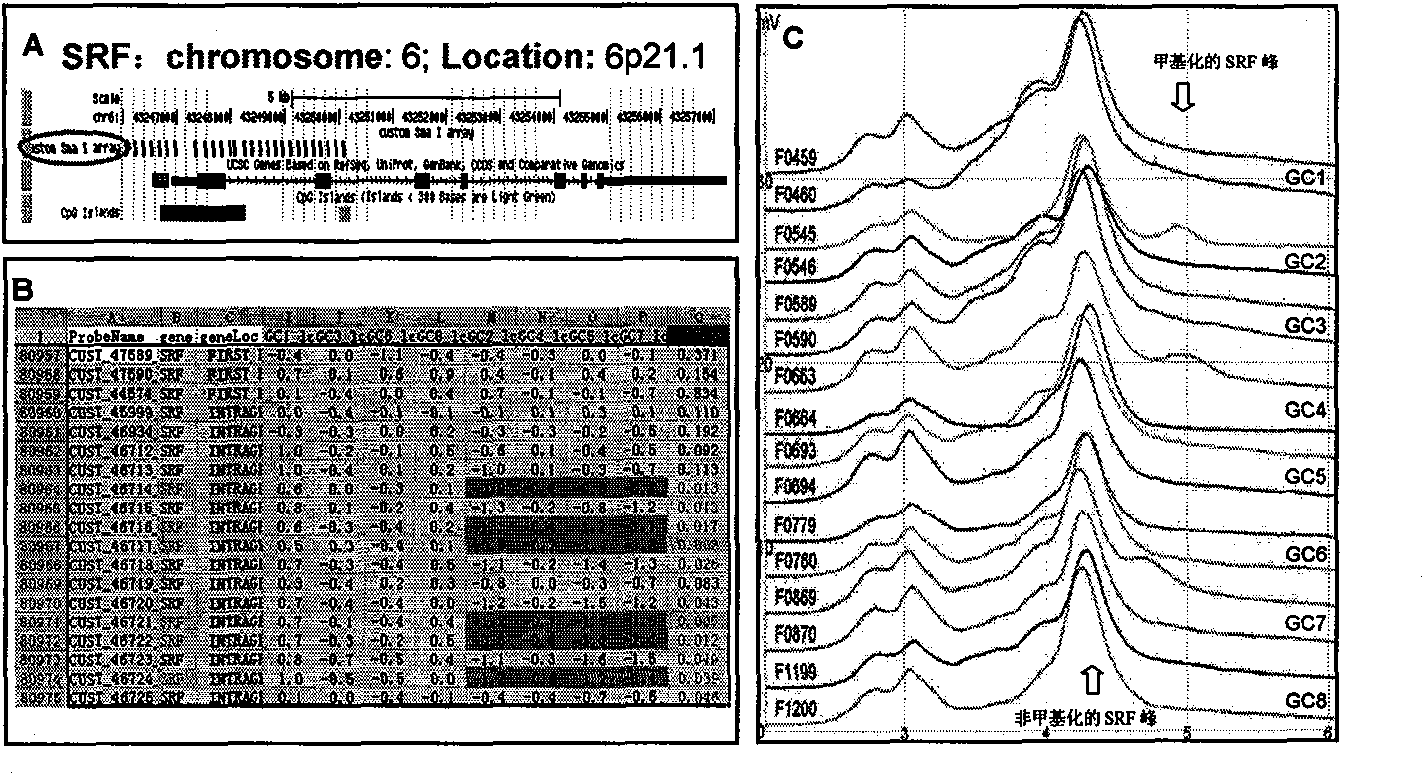
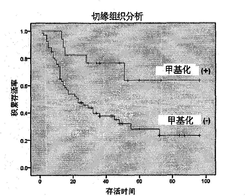
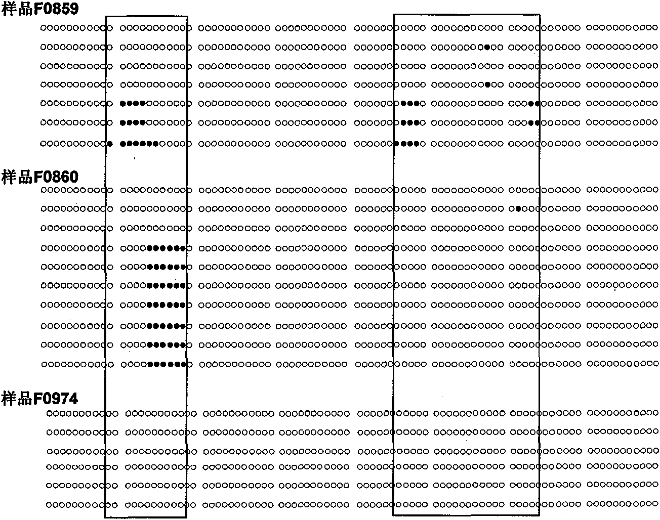
[0094] Example 1: Predicting the ability of gastric cancer invasion and metastasis by determining whether there is methylation of cytosine in the CpG island in the SRF nucleotide sequence in gastric cancer margin tissue

[0095]1. Experimental subjects: 102 paired frozen tissues of gastric cancer and their surgical margins, 50% of the patients had lymph node metastases and vascular tumor thrombi (or distant metastases), and 50% of the paired gastric cancer tissues had no lymph node metastasis No vascular tumor thrombus (or distant metastases) was seen, and complete clinicopathological data and follow-up data were available;

[0096] 2. Obtain tissue cells: cut out the size of rice grains (15-20 mm) from different parts of the surgical margin tissue 3 ) of five frozen tissues were collected into siliconized microcentrifuge tubes (1.5ml);

[0097] 3. Extract cell DNA: add 300μl tissue cell lysate (containing 10mM Tris.Cl / pH 7.6, 10mM NaCl, 10mM EDTA and 0.5% SDS) to the centrif...

Embodiment 2

[0114] Example 2: Predicting the ability of invasion and metastasis of thyroid cancer by determining whether there is methylation of cytosine in the CpG island in the SRF nucleotide sequence in the resection margin tissue of thyroid cancer

[0115] 1. Subjects: 20 pairs of thyroid cancer frozen tissues, 50% of the patients had lymph node metastasis and vascular tumor thrombus (or distant metastasis), and 50% of the paired patients had neither lymph node metastasis nor vascular cancer Thrombus (or distant metastases), all have complete clinicopathological data and follow-up data;

[0116] 2. Steps 2-7 are the same as in Example 1;

[0117] 3. Results ( Figure 4 ) and conclusion are the same as in Example 1.

Embodiment 3

[0118] Example 3: Prediction of gastric cancer invasion and metastasis by determining whether there is methylation of cytosine in CpG island in SRF nucleotide sequence in gastric cancer margin tissue

[0119] Steps 1-5 are the same as in Example 1; in step 6, except for determining whether there is a serum response factor gene DNA sequence whose number of methylated cytosines in the 5'-terminal CpG island is greater than or equal to 6, the rest are the same as in Example 1 Step 6 in is identical; The specificity of the measured result is higher than embodiment 1, is 100%.


PUM


Abstract

The invention discloses a method for predicting malignant tumor metastasis and invasion capacity in vitro, which comprises the following steps of: (a) extracting DNA (Deoxyribonucleic Acid) from surgical incisal margins or tumor-adjacent tissues; (b) detecting the methylating degree of cytimidine in 5'- end CpG island of seroreaction factor gene DNA sequence in DNA obtained in the step (a); and (c) when the seroreaction factor gene DNA sequence with methylated cytimidine in the 5'-end CpG island is detected in the step (b), presuming that the tissue being detected has the capacity of resisting malignant tumor metastasis and invasion. The invention also provides four artificial nucleotide fragments. The inventor of the invention, for the first time, find that the metastasis and invasion capacity of malignant tumors can be rapidly and accurately predicted by detecting whether the seroreaction factor gene DNA sequence with methylated cytimidine in CpG island exists or not in the tissue being detected. Besides, the nucleotide fragments provided in the invention can be used for accurately and rapidly detecting the methylation of cytimidine in the CpG island in the seroreaction factor gene DNA sequence.

Description

technical field [0001] The invention relates to a method for predicting the invasion and metastasis ability of malignant tumors in vitro and the artificial nucleotide fragments used. Background technique [0002] The ability to invade and metastasize malignant tumors is an important factor in determining the prognosis of patients. Tumor tissue invasion and destruction of adjacent organs and distant metastasis are not only the main factors that make patients lose the chance of surgical treatment, but also the basic reason for postoperative recurrence leading to treatment failure. Establishing a method to predict the invasion and metastasis of malignant tumors is a long-term pursuit of people, and it has important guiding value for formulating treatment plans for tumor patients after surgery. [0003] It is difficult to judge the metastasis and recurrence of patients after surgery according to the pathological changes of tumors. People have high hopes for the molecular classi...

Claims


Application Information

Patent Timeline
IPC IPC(8): C12Q1/68C12N15/11
CPCC12Q2600/154C12Q2600/118C12Q1/6886
Inventor 邓大君刘兆君
Owner BEIJING CANCER HOSPITAL PEKING UNIV CANCER HOSPITAL
Why Eureka
Try Eureka