
Template-based CCD-DR (charge coupled device-digital radiography) image splicing method

A CCD-DR, image stitching technology, applied in the field of medical image stitching, can solve the problems of image chromatic aberration, inability to obtain high-quality medical stitching images, artifact stitching efficiency, etc.

Inactive Publication Date: 2011-09-14
NANCHANG HANGKONG UNIVERSITY
View PDF2 Cites 19 Cited by
  • Summary
  • Abstract
  • Description
  • Claims
  • Application Information

AI Technical Summary

Problems solved by technology

However, in view of the single particularity of the orthopedic medical image scene, high-quality medical stitching images cannot be obtained by using the traditional stitching and fusion process technology. Defects such as image chromatic aberration and artifacts are common and the stitching efficiency is low.

Method used


Image

  • Template-based CCD-DR (charge coupled device-digital radiography) image splicing method

Examples


Embodiment Construction

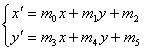
[0016] The method of the present invention requires the use of a CCD array to collect DR images within a certain field of view for image splicing, and first uses the grid template DR images to determine the position transformation relationship of adjacent CCD-DR images, and maintains stability under the CCD-DR image acquisition conditions Under the same conditions, the saved grid template DR image registration information can be repeatedly called to stitch the real DR image.

[0017] The flowchart of the template-based CCD-DR image mosaic method of the present invention obtains a panoramic DR image with high resolution and large field of view, and its specific implementation steps are as follows:

[0018] 1. CCD-DR image acquisition

[0019] The CCD array is used to collect the equal-spaced circular lattice template and the real DR image respectively, and the image data is input into the computer system.

[0020] 2. CCD-DR image preprocessing

[0021] The acquired CCD-DR ima...


PUM


Abstract

The invention provides a template-based CCD-DR (charge coupled device-digital radiography) image splicing method, comprising the following steps of: 1) collecting the template and a real object DR image in a certain visual field range by using a CCD array; 2) pre-processing the acquired CCD-DR image; 3) extracting characteristic points from the template DR images; 4) performing registration to the characteristic points of the adjacent template DR images; and 5) splicing a plurality of real object DR images which are collected by the CCD array according to the registration relationship of characteristic points acquired in the step 4). The method performs registration to the characteristic points of the template DR images, determines the position change relationship between the adjacent DR images in the CCD array and stores the related image registration information; under the condition that the CCD-DR image collection conditions are unchanged, the image registration information is repetitively transferred to splice the real object DR images so as to highly-efficiently acquire panoramic DR images with high resolution and large visual field.

Description

technical field [0001] The invention relates to a medical image mosaic method, that is, to a method for collecting DR images within a certain field of view by using a CCD array, and using the positional relationship determined by the template CCD-DR image to realize the mosaic method of the real CCD-DR image. Background technique [0002] With the continuous development of visualization technology and modern medical technology, X-ray medical images are widely used in medical image analysis such as clinical diagnosis and medical research. Especially in image-guided orthopedic diagnosis and treatment, it has important clinical application value. In the orthopedic diagnosis and surgical treatment of the human spine and long bones, it is often necessary to obtain large-scale, super-resolution, and complete anatomical structure images to conduct comprehensive and intuitive observation and analysis of the patient's lesion and its surrounding parts. [0003] At present, digital X-...

Claims


Application Information

Patent Timeline
IPC IPC(8): G06T11/60G06K9/46G06K9/36A61B6/00
Inventor 熊邦书陈乐平欧巧凤
Owner NANCHANG HANGKONG UNIVERSITY
Learn More