Time intensity characteristic-based computer aided method for diagnosing benign and malignant breast lesions

A computer-aided, time-intensive technology, applied in diagnosis, calculation, diagnostic recording/measurement, etc., can solve the problems of missed or misdiagnosed lesions, increased radiologist reading pressure, and difficulty in ensuring the accuracy and validity of lesion diagnosis. Achieve the effect of improving diagnostic efficiency and accuracy, and improving diagnostic efficiency and accuracy

Inactive Publication Date: 2011-11-23
DALIAN UNIV OF TECH
View PDF2 Cites 14 Cited by
  • Summary
  • Abstract
  • Description
  • Claims
  • Application Information

AI Technical Summary

Problems solved by technology

However, multi-sequence DCE-MRI images increase the reading pressure of radiologists
Subjective factors such as visual fatigue of doctors caused by this may cause missed or misdiagnosed lesions, which makes it difficult to guarantee the accuracy and effectiveness of lesions diagnosis

Method used


Image

  • Time intensity characteristic-based computer aided method for diagnosing benign and malignant breast lesions
  • Time intensity characteristic-based computer aided method for diagnosing benign and malignant breast lesions
  • Time intensity characteristic-based computer aided method for diagnosing benign and malignant breast lesions

Examples


Embodiment Construction

[0025] The present invention will be described in further detail below in conjunction with accompanying drawings and examples.

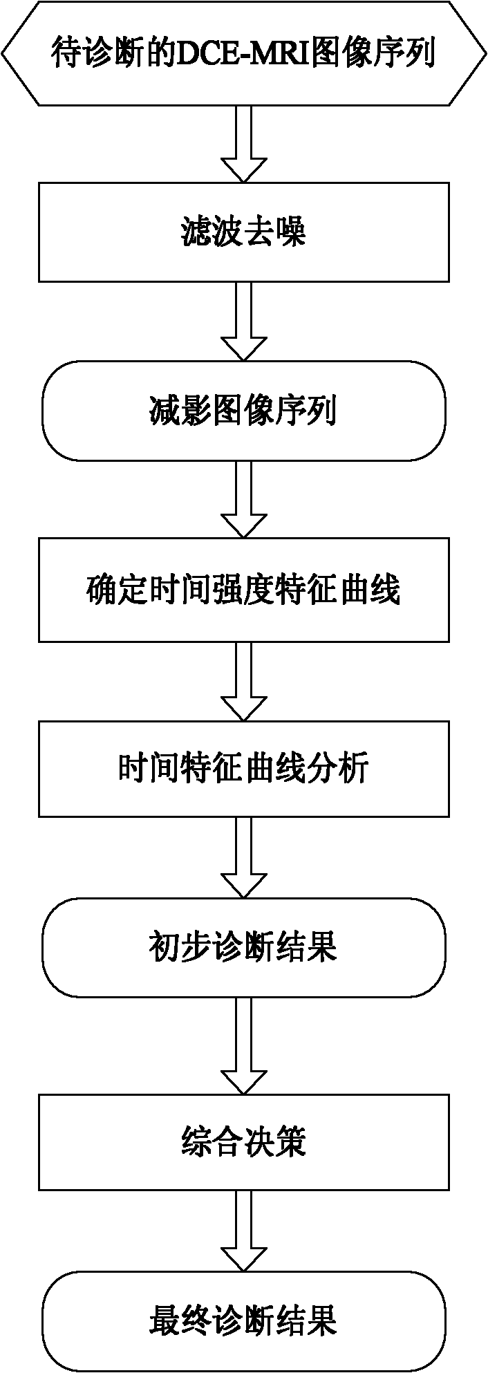
[0026] Such as figure 1 , 2 As shown, the inventive method comprises the following processing steps:

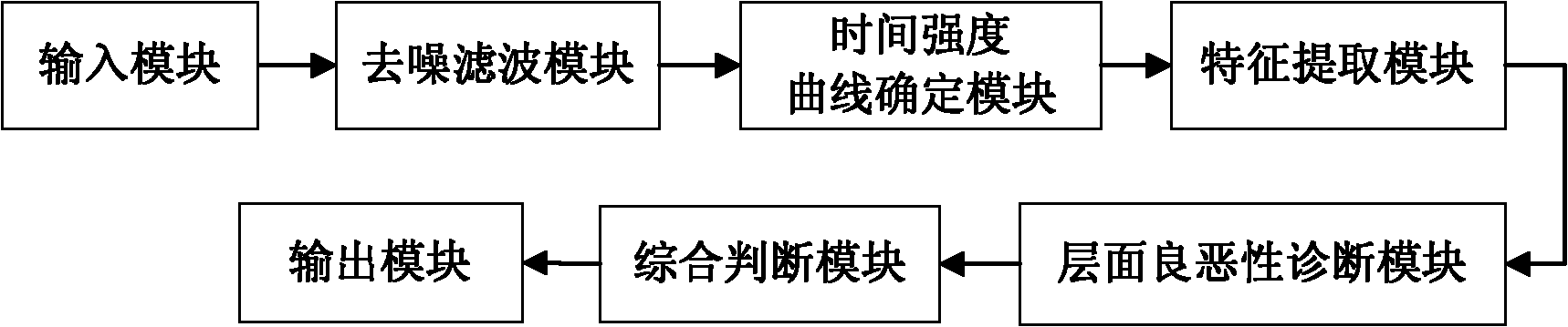
[0027] (1) In the input module, select the suspicious layer image sequence in the patient's DCE-MRI image set according to the scanning order;

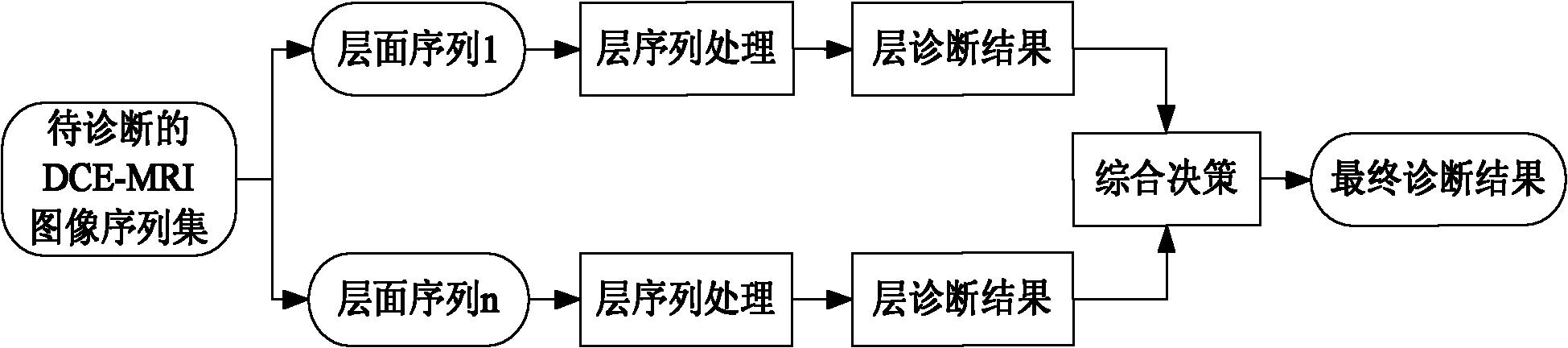
[0028] DCE-MRI image data is a multi-sequence, multi-temporal image set. Multi-sequence can obtain the complete spatial information of the lesion, while multi-temporal phase can observe the intensity changes at different times at the same level and provide the physiological and metabolic information of the lesion at this level. Such as image 3 As shown, the processing sequence of image data among the present invention is: first according to Figure 4 The processing steps shown in analyze the phase information of each layer in the sequence image set of suspicious layers, and then combine the diagnostic...


PUM


Abstract

The invention discloses a time intensity characteristic-based computer aided method for diagnosing benign and malignant breast lesions, comprising the following steps of: selecting an image sequence layer with suspicious lesions from a DCE(Dynamic Contrast-Enhanced)-MRI (Magnetic Resonance Imaging) image sequence set; carrying out denoising and filtering treatment on each image layer and acquiring a photographic subtraction sequence of the layer; determining a time intensity curve according to the photographic subtraction sequence of the layer; analyzing the characteristic of the time intensity curve and giving out a diagnosis result of the layer, namely a preliminary diagnosis result of the lesion; and fusing diagnosis results of the time intensity curves for different layers to give out a final lesion diagnosis result. According to the time intensity characteristic-based computer aided method disclosed by the invention, by comprehensively analyzing the characteristic of the time intensity curve on each layer, the accuracy of benign and malignant diagnosis of the lesions can be greatly improved for assisting clinical diagnosis for breast diseases and further the misdiagnosis rate is reduced.

Description

technical field [0001] The present invention relates to the field of computer-aided diagnosis of breast diseases. More specifically, the present invention relates to a computer-aided method for benign and malignant breast lesions based on DCE-MRI image processing, time-intensity curve feature extraction, classification and decision fusion. Background technique [0002] Breast cancer is one of the most common malignant tumors in women. According to statistics, in recent years, the incidence of breast cancer in my country has continued to rise significantly. If breast cancer can be detected, diagnosed and treated early, the cure rate and patient survival rate of breast cancer can be improved. As a non-invasive examination technique, MRI technology is the most potential imaging examination method for diagnosing breast diseases at this stage, which has been fully reflected in clinical applications. MRI technology can provide multi-parameter, multi-sequence, multi-directional i...

Claims


Application Information

Patent Timeline
Patent Type & Authority Applications(China)
IPC IPC(8): A61B5/055G01R33/56G06F19/00
Inventor 刘惠李珂
Owner DALIAN UNIV OF TECH
Why Eureka
Try Eureka