Device and method for generating soft tissue contrast images
A soft tissue, contrast technique used in instruments for radiological diagnosis, material analysis using radiation diffraction, applications, etc.
- Summary
- Abstract
- Description
- Claims
- Application Information
AI Technical Summary
Problems solved by technology
Method used
Image
Examples
Embodiment Construction
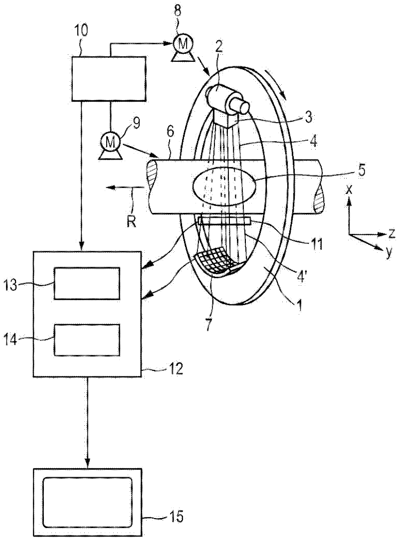
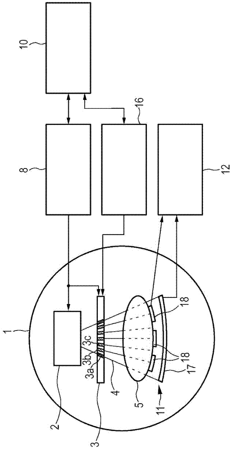
[0045] figure 1 A first embodiment of the device according to the invention is shown. In this embodiment, the mechanical layout is similar to conventional CT imaging devices. The device comprises a gantry 1 which is rotatable about an axis of rotation R extending parallel to the z-direction. A radiation source unit including an X-ray source 2 such as an X-ray tube is mounted on the gantry 1 . The X-ray source unit further comprises a collimator unit 3 which forms a pencil beam 4 (at least one pencil beam) from the radiation beam emitted by the X-ray source 2 .
[0046] The pencil beam penetrates an object 5 (shown symbolically), such as a patient, in a region of interest within the cylindrical examination region 6 . After penetrating the examination region 6, the part of the pencil beam's X-ray radiation 4' that is not absorbed by the object 5 is incident on the X-ray detector unit 7, which in this embodiment is a two-dimensional An energy-resolving detector, which is also...
PUM
| Property | Measurement | Unit |
|---|---|---|
| current density | aaaaa | aaaaa |
| energy | aaaaa | aaaaa |
Abstract
Description
Claims
Application Information
 - R&D
- Intellectual Property
- Life Sciences
- Materials
- Tech Scout
- Unparalleled Data Quality
- Higher Quality Content
- 60% Fewer Hallucinations
Browse by: Latest US Patents, China's latest patents, Technical Efficacy Thesaurus, Application Domain, Technology Topic, Popular Technical Reports.
© 2025 PatSnap. All rights reserved.Legal|Privacy policy|Modern Slavery Act Transparency Statement|Sitemap|About US| Contact US: help@patsnap.com


