Ultrasonic scanning tomography device for volumes of superficial tissues and organs

A technology of scanning tomography and imaging equipment, applied in the direction of echo tomography, etc., can solve the problems of not being suitable for widespread promotion, lack of flexibility, and high cost of a complete set of equipment, and achieve the effects of portability, simplified requirements, and miniaturization

Inactive Publication Date: 2012-10-03
HUADONG HOSPITAL
View PDF4 Cites 16 Cited by
  • Summary
  • Abstract
  • Description
  • Claims
  • Application Information

AI Technical Summary

Problems solved by technology

Their common disadvantage is that the application flexibility is insufficient, and the cost of a complete set

Method used


Image

  • Ultrasonic scanning tomography device for volumes of superficial tissues and organs
  • Ultrasonic scanning tomography device for volumes of superficial tissues and organs
  • Ultrasonic scanning tomography device for volumes of superficial tissues and organs

Examples


Embodiment Construction

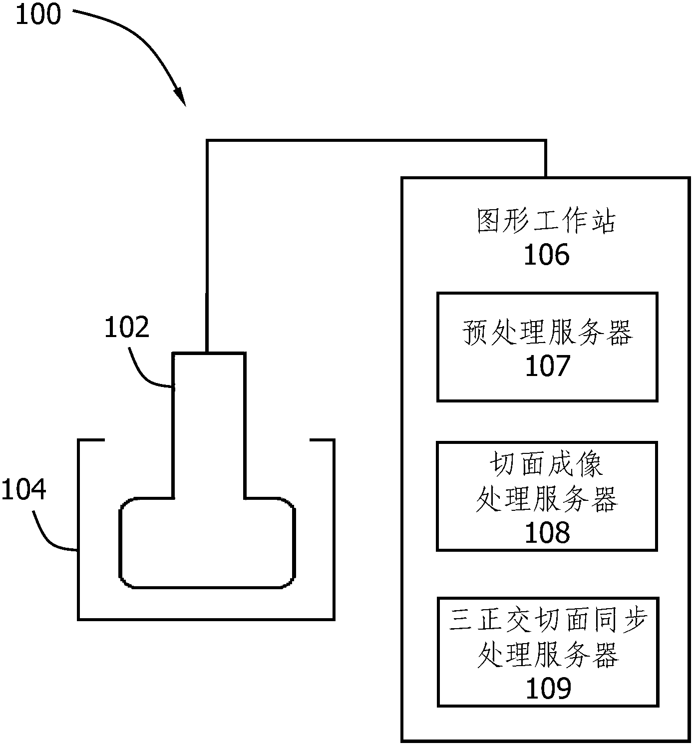
[0024] refer to figure 1 as shown, figure 1 A structural diagram of an ultrasonic superficial tissue and organ volume imaging device according to an embodiment of the present invention is disclosed. The ultrasonic superficial tissue and organ volume imaging device 100 includes: a probe 102 , a bracket 104 and a graphics workstation 106 . The probe 102 emits ultrasonic waves and receives echoes. The probe 102 has a concave end, and the probe 102 collects continuous multi-frame two-dimensional slice ultrasonic image data of superficial tissues and organs. The probe 102 is installed on the bracket 104, the bracket 104 drives the probe to move and generates a trigger signal, and the probe 102 samples a sliced ​​two-dimensional ultrasound image according to the trigger signal. The graphics workstation 106 is connected to the probe 102, the graphics workstation 106 acquires the two-dimensional ultrasound image generated by the probe 102, and reconstructs the cross-section, sagitta...


PUM

PropertyMeasurementUnit
Lengthaaaaaaaaaa

Abstract

The invention discloses an ultrasonic scanning tomography device for volumes of superficial tissues and organs, which comprises a probe, a bracket and a graphic workstation, wherein the probe is provided with an indent end part and is used for emitting an ultrasonic wave, receiving an echo and collecting two-dimensional ultrasonic images of the superficial tissues and organs along the direction vertical to the body surface; the probe is mounted on the bracket; the bracket is used for driving the probe to move and generating a trigger signal; the continuous multi-frame two-dimensional ultrasonic images are sampled by the probe according to the trigger signal; the graphic workstation is connected with the probe and is used for obtaining volume data of the two-dimensional ultrasonic images and reconstructing the images of three orthogonal tangent planes, namely, a cross tangent plane, a vertical plane and a coronal plane of the superficial tissues and organs, on the basis of the two-dimensional ultrasonic images; and the graphic workstation comprises a preprocessing server, a tangent plane image processing server and a synchronous processing server for the three orthogonal tangent planes. The probe with the indent end part adopted by the ultrasonic scanning tomography device provided by the invention is suitable for the detection for the superficial tissues and organs, such as mammary glands, thyroid glands, four limbs and the trunk. The process of imaging the three orthogonal tangent planes of the graphic workstation is simpler.

Description

technical field [0001] The invention relates to the field of medical imaging equipment, in particular to a volume imaging equipment for superficial tissues and organs. Background technique [0002] Ultrasound (Ultrasound) medicine is a discipline that combines acoustics, medicine, optics and electronics. Ultrasound medicine is the study of the application of acoustic technology higher than audible sound frequency in the medical field. Including ultrasonic diagnostics, ultrasonic therapeutics and biomedical ultrasonic engineering, etc., so ultrasonic medicine has the characteristics of combining medicine, science and engineering, involves a wide range of contents, and has high value in the prevention, diagnosis and treatment of diseases. [0003] An important item in ultrasound medicine is ultrasound imaging technology. Ultrasound imaging technology is to transmit ultrasonic waves by a probe and receive the reflected ultrasonic waves. An image is generated based on the ech...

Claims


Application Information

Patent Timeline
IPC IPC(8): A61B8/14
Inventor 陈悦陈林
Owner HUADONG HOSPITAL
Why Eureka
Try Eureka