S-shaped visible hard cannula core

A rigid tube and visual technology, applied in the field of medical devices, can solve the problems that clinicians cannot accurately control the image taking direction of the micro camera, lack of rigidity, etc., and achieve the effect of convenient operation, good accuracy and shortened operation time for doctors

Active Publication Date: 2012-10-31
ZHEJIANG UE MEDICAL
View PDF5 Cites 5 Cited by
  • Summary
  • Abstract
  • Description
  • Claims
  • Application Information

AI Technical Summary

Problems solved by technology

[0005] The present invention mainly solves the technical problem that clinicians cannot accurately control the image-taking direction of the micro-camera due to insufficient rigidity of the original bendable optical fiber or video bronchosc

Method used


Image

  • S-shaped visible hard cannula core
  • S-shaped visible hard cannula core
  • S-shaped visible hard cannula core

Examples


Example Embodiment

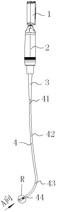
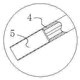
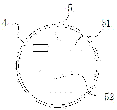
[0026] Embodiment: a kind of S-shaped visible hard intubation core, as figure 1 As shown, it includes a liquid crystal display 1, a hollow handle 2, a hollow endotracheal tube positioner 3 and a hollow hard tube core 4, one end of the handle 2 is fixedly connected with the liquid crystal display 1, and the other end of the handle 2 is connected with the endotracheal tube One end of the locator 3 is threadedly connected, and the other end of the endotracheal catheter locator 3 is closely matched with one end of the hard tube core 4, and the other end of the hard tube core 4 is provided with a miniature camera 5 and an LED light 51, the miniature camera 5 and LED lights 51 are respectively connected with the liquid crystal display 1, and the hard tube core 4 from one end to the other end is respectively the first linear section 41, the arc section 42, the arc section 43, and the second straight section 44. , the arc-shaped section 42 and the arc-shaped section 43 are in the shap...


PUM

PropertyMeasurementUnit
Arc radiusaaaaaaaaaa
Central angleaaaaaaaaaa
Angleaaaaaaaaaa

Abstract

The invention discloses an S-shaped visible hard cannula core. The S-shaped visible hard cannula core comprises a liquid crystal display, a handle and a hard tube core which are sequentially connected, wherein a camera and a light-emitting body are arranged at the outer end of the hard tube core and respectively connected with the liquid crystal display. The hard tube core is sequentially divided into a first linear section, a camber-line-shaped section, a circular arc section and a second linear section from one end to the other end, wherein the included angle formed by the tangential direction of the tail end of the circular arc section close to the camber-line-shaped section and the axis direction of the handle ranges from 2 degrees to 20 degrees, the circular arc radius of the circular arc section is 45-70mm, the central angle which the circular arc of the circular arc section faces ranges from 60 degrees to 80 degrees, and the included angle formed by the second linear section and the axis direction of the handle ranges from 50 degrees to 70 degrees. The S-shaped visible hard cannula core has the advantages of being convenient to operate, clear in images and capable of fast and accurately finishing trachea cannula inserting operation.

Description

technical field [0001] The invention belongs to the technical field of medical devices, and in particular relates to a visible hard intubation core used for guiding a patient's tracheal intubation. Background technique [0002] The laryngoscope, which is a necessary instrument for tracheal intubation in the anesthesiology department, emergency department and intensive care department of the hospital, has been widely used in clinical practice. At present, the laryngoscope with video function usually inserts the endotracheal tube with the intubation core directly into the patient's trachea under the video image provided by the camera, or uses the guide groove designed on one side of the laryngoscope to insert the endotracheal tube into the patient's trachea. The catheter is inserted into the patient's trachea along the guide groove under the guidance of the video image, such as a document CN201328799Y published by the Chinese Patent Office on October 21, 2009. The name of the ...

Claims


Application Information

Patent Timeline
IPC IPC(8): A61M16/04
Inventor 薛富善马武华陈爱华杨本全王卫东李岳胜赵燕勇朱福斌周旭林张文銮
Owner ZHEJIANG UE MEDICAL
Why Eureka
Try Eureka