Tube for inspecting cervix uteri including hollow part for monitoring and detector including same

A cervix and detector technology, applied in the field of detectors, can solve the problems of inconvenience, pain, and unwillingness of patients, and achieve the effect of improving patient satisfaction, accurate diagnosis, and reducing the probability of infection

Active Publication Date: 2012-11-21
首尔大学校病院 分事务所
View PDF7 Cites 0 Cited by
  • Summary
  • Abstract
  • Description
  • Claims
  • Application Information

AI Technical Summary

Problems solved by technology

At this time, due to the insertion of the doctor's hand or speculum, the narrow vaginal entrance is forced to open, which makes the patient feel inconvenient or painful, so there is a problem that the patient is unwilling to accept these examinations, and even refuses the examination in severe cases

Method used


Image

  • Tube for inspecting cervix uteri including hollow part for monitoring and detector including same
  • Tube for inspecting cervix uteri including hollow part for monitoring and detector including same
  • Tube for inspecting cervix uteri including hollow part for monitoring and detector including same

Examples


Embodiment 1

[0064] Example 1: Structure of Catheter and Detector for Cervical Examination

[0065] 1-1. Structure of catheter for cervical examination

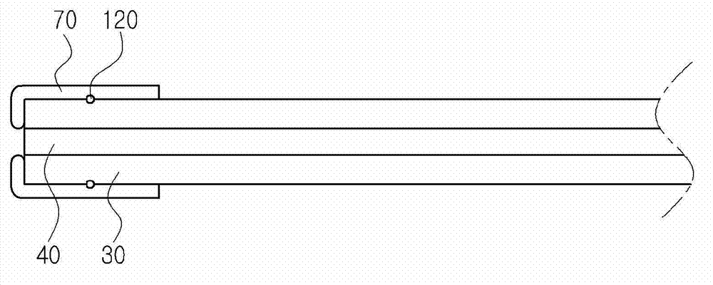
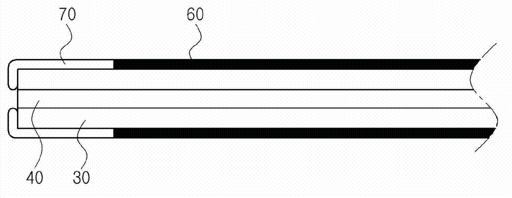
[0066] Such as Figure 2 to Figure 7 As shown, the catheter for cervical examination of the present invention includes: a hollow catheter 30, which is inserted into the vagina 10; a volume expansion part 70, which is arranged at one end of the catheter, so that a hollow part for monitoring is formed when the volume is expanded; And a volume control part 100 configured to expand the volume of the volume expansion part 70 .

[0067] As a specific example, the catheter for cervical examination of the present invention may be disposable.

[0068] One end of the conduit can pass through to form a medium inlet and outlet hole 120, so that the volume medium supplied to the volume control part 100 flows into the side of the volume expansion part 70 ( figure 2 ).

[0069] As a specific example, the volume expansion part 70 may use a balloon...

Embodiment 2

[0087] Example 2: Role of Catheters and Detectors for Cervical Examination

[0088] The detector for cervical examination of the present invention is inserted into the vagina 10 . When reaching the cervix 20 through the entrance of the vagina 10 and the upper part of the vagina 10, the volume of the volume expansion part 70 will be expanded by the volume control part 100, so that the lower part of the vagina 10 in a collapsed state will be opened, thereby ensuring the front part of the cervix 20 Space. The state of the cervix 20 can be directly confirmed by the monitor through the lens of the cervix detecting portion 50 . Patients can also confirm their own cervix20 through the monitor.

[0089] Furthermore, by measuring the size of the entrance (os) of the cervix 20 with the monitor, the progressing stage of labor pains can be grasped.

[0090] In addition, amniotic fluid or vaginal secretions can be sucked through the suction part additionally provided in the detector f...


PUM


Abstract

The present invention provides a tube for inspecting cervix uteri and comprising: a hollow tube (30) which is inserted into the vagina (10); a volume expanding part (70) which is installed at one end of the tube including the hollowing part for monitoring the cervix uteri (20) at the time of volume expansion; and a volume control part (100) which is prepared for expanding volume of the volume expanding part (70). In addition, the present invention provides a detector for inspecting the cervix uteri including the tube for inspecting the cervix uteri and a detecting part (50) of the cervix uteri which is placed by passing through the tube. The tube for inspecting the cervix uteri and the detector of the present invention can eliminate inconvenience and pain of obstetric patients, and effectively inspects the cervix uteri widening the lower part of the vagina at the front of the cervix uteri without widening the upper part and entrance thereof. In addition, the change of cervix uteri of a woman being in labor can accurately and continuously be taken without vaginal examination, and more objective and accurate judgment can be obtained for the requirement of performing a cesarean. Since the woman in childbed confirms the change of the cervix uteri on the monitor, the bio-feedback effect can be expected. Furthermore, it is able to accurately confirm early contraction or cervix uteri condition of a patient of premature rupture of membrane before the last month of pregnancy for more precise diagnosis and decrease of infection chance. In addition, the contentment of the patient can be enhanced since obstetrics inspection with explanation can be performed without a speculum at ambulatory care directly showing the cervix uteri condition to the patient.

Description

technical field [0001] The present invention relates to a catheter for cervical examination including a hollow portion for monitoring and a detector including the catheter. Background technique [0002] Knowing the status of the cervix is ​​very important for the diagnosis and treatment of obstetrics and gynecology, and as a basic examination, it is an examination that is required for almost all patients. To do this, the doctor puts a hand directly into the vagina and touches the cervix (palpation) to understand its condition (internal examination) or puts a speculum (speculum) into the vagina to open the vagina and checks the cervix with eyes (inspection) , inspection)( figure 1 ). At this time, due to the insertion of the doctor's hand or speculum, the narrow vaginal entrance is forced to open, which makes the patient feel inconvenient or painful, so there is a problem that the patient is unwilling to accept these examinations, and even refuses the examination in severe ...

Claims


Application Information

Patent Timeline
Patent Type & Authority Applications(China)
IPC IPC(8): A61B1/303
CPCA61B1/0008A61B1/303A61B1/00082
Inventor 尹保铉洪焌硕
Owner 首尔大学校病院 分事务所
Why Eureka
Try Eureka