Cavity body inside three-dimensional ultrasound probe used in ultrasound medical equipment

A medical equipment, three-dimensional ultrasound technology, applied in the direction of catheter, surgery, etc., can solve the problem of inconsistency in intracavity images, etc., to achieve fast and accurate processing and real-time display, reliable structure, and uniform data collection

Inactive Publication Date: 2013-06-26
SHENZHEN WELL D MEDICAL ELECTRONICS
View PDF8 Cites 0 Cited by
  • Summary
  • Abstract
  • Description
  • Claims
  • Application Information

AI Technical Summary

Problems solved by technology

This requires higher requirements for the doctor or operator's operation techniques, and different operators may present inconsistent intracavity images by using this method

Method used


Image

  • Cavity body inside three-dimensional ultrasound probe used in ultrasound medical equipment
  • Cavity body inside three-dimensional ultrasound probe used in ultrasound medical equipment
  • Cavity body inside three-dimensional ultrasound probe used in ultrasound medical equipment

Examples


Embodiment Construction

[0018] In order to fully understand the technical content of the present invention, the technical solution of the present invention will be further introduced and illustrated below in conjunction with specific embodiments.

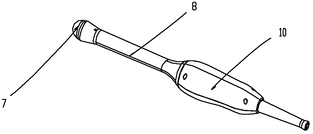
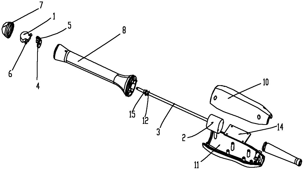
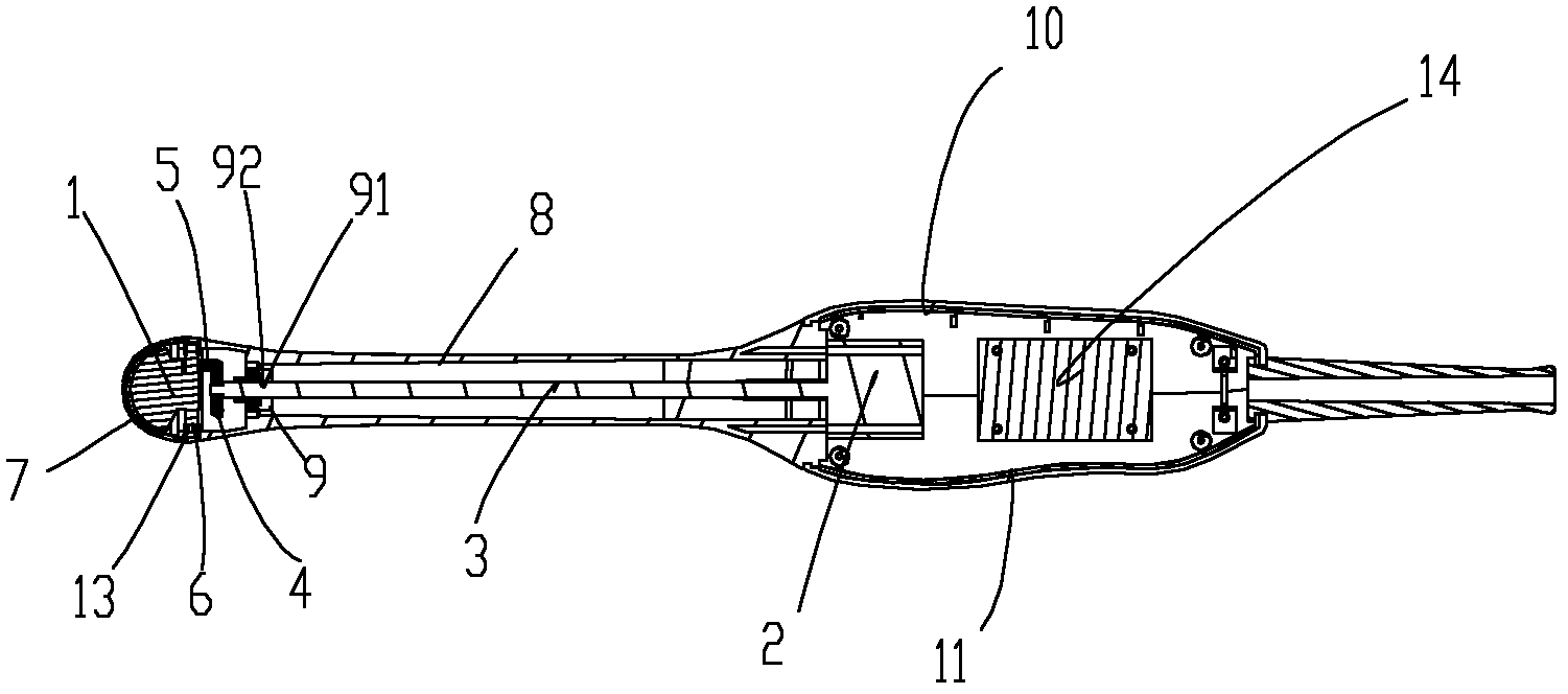
[0019] Such as figure 1 As shown, it is a schematic diagram of the three-dimensional structure of the present invention; as figure 2 Shown is a schematic diagram of the explosion structure of the present invention; as image 3 As shown, it is a cross-sectional view of the present invention, including an acoustic head 1 and an outer casing that seals the sound head, and a driving motor 2 electrically connected to the motor drive control device in the ultrasonic medical equipment is arranged in the outer casing, and the driving motor 2 The output shaft 3 is provided with a driving bevel gear 4, the sound head 1 is fixed on a rotating shaft 6 with a driven bevel gear 5, and the driving bevel gear 4 and the driven bevel gear 5 are meshed for transmission . ...


PUM

PropertyMeasurementUnit
Lengthaaaaaaaaaa

Abstract

The invention relates to a cavity body inside three-dimensional ultrasound probe used in ultrasound medical equipment. The cavity body inside three-dimensional ultrasound probe used in the ultrasound medical equipment comprises a sound head and a shell body, wherein the sound head is sealed by the shell body. A driving motor which is in electric connection with a motor drive control device of the ultrasound medical equipment is arranged in the shell body. An active umbrella-shaped gear is arranged on an output shaft of the driving motor. The sound head is fixedly on a rotating shaft with a passive umbrella-shaped gear. The active umbrella-shaped gear and the passive umbrella-shaped gear are meshed to drive. A multi array element sound head can automatically and rapidly conduct three-dimensional scanning the cavity body inside under control of the medical equipment, collection of ultrasound real-time three-dimensional data on the scanning portion can be achieved automatically, and the data collection has the advantages of being even, continuous and rapid, convenient and reliable, and reliable in structure, and rapid and accurate treatment and real-time display of ultrasound three-dimensional images through the medical equipment are guaranteed.

Description

technical field [0001] The invention relates to a medical device, in particular to an in-cavity three-dimensional ultrasonic probe for an ultrasonic medical device. Background technique [0002] The current three-dimensional ultrasound technology in China is still limited to the outside body, and can only display images through bulky three-dimensional probes, and there is no intracavity three-dimensional ultrasound probe. At present, to achieve all-round image observation in the cavity, it mainly relies on the ordinary two-dimensional intracavity probe and realizes it by changing different scanning methods. This requires higher requirements on the operation techniques of doctors or operators, and different operators may present intracavity images with inconsistent effects by using this method. The true three-dimensional intracavity ultrasound probe solves this problem, and it is not limited by the scanning method. Doctors only need to place the probe, and then they can view...

Claims


Application Information

Patent Timeline
IPC IPC(8): A61B8/12
Inventor 黄小划毛志林白宁全宏岳李元强李青松詹凯刘立元
Owner SHENZHEN WELL D MEDICAL ELECTRONICS
Why Eureka
Try Eureka