Extension ultrasound vascular imaging method and device based on catheter path

A vascular imaging and vascular technology, applied in the field of surgical navigation, can solve problems such as lack of depth information, kidney damage, and patient allergies

Inactive Publication Date: 2013-09-11
HARBIN ENG UNIV
View PDF6 Cites 22 Cited by
  • Summary
  • Abstract
  • Description
  • Claims
  • Application Information

AI Technical Summary

Problems solved by technology

This method has the following disadvantages: First, X-ray perspective imaging equipment can only produce two-dimensional projection images, lacking depth information
It is difficult for the operator to make the guide wire reach the target position quickly and accurately under the 2D projection image
Wrong or repeated operations may cause certain damage to human blood vessels, which brings certain risks to the operation
Secondly, the use of intraoperative X-ray images causes medical staff and patients to be exposed to large doses of radiation, which brings varying degrees of damage to physical and mental health and increases the risk of cancer
Third, because vascular imaging requires the use of special contrast agents, large doses of contrast agents can cause allergies in patients and damage the kidneys.
However, these methods require the use of complete ultrasound images. For blood vessels in areas that cannot be imaged by ultrasound, it is impossible to accurately obtain images of blood vessels due to possible deformation of the blood vessels.

Method used


Image

  • Extension ultrasound vascular imaging method and device based on catheter path
  • Extension ultrasound vascular imaging method and device based on catheter path
  • Extension ultrasound vascular imaging method and device based on catheter path

Examples


Embodiment Construction

[0024] The present invention will be further described below in conjunction with accompanying drawing:

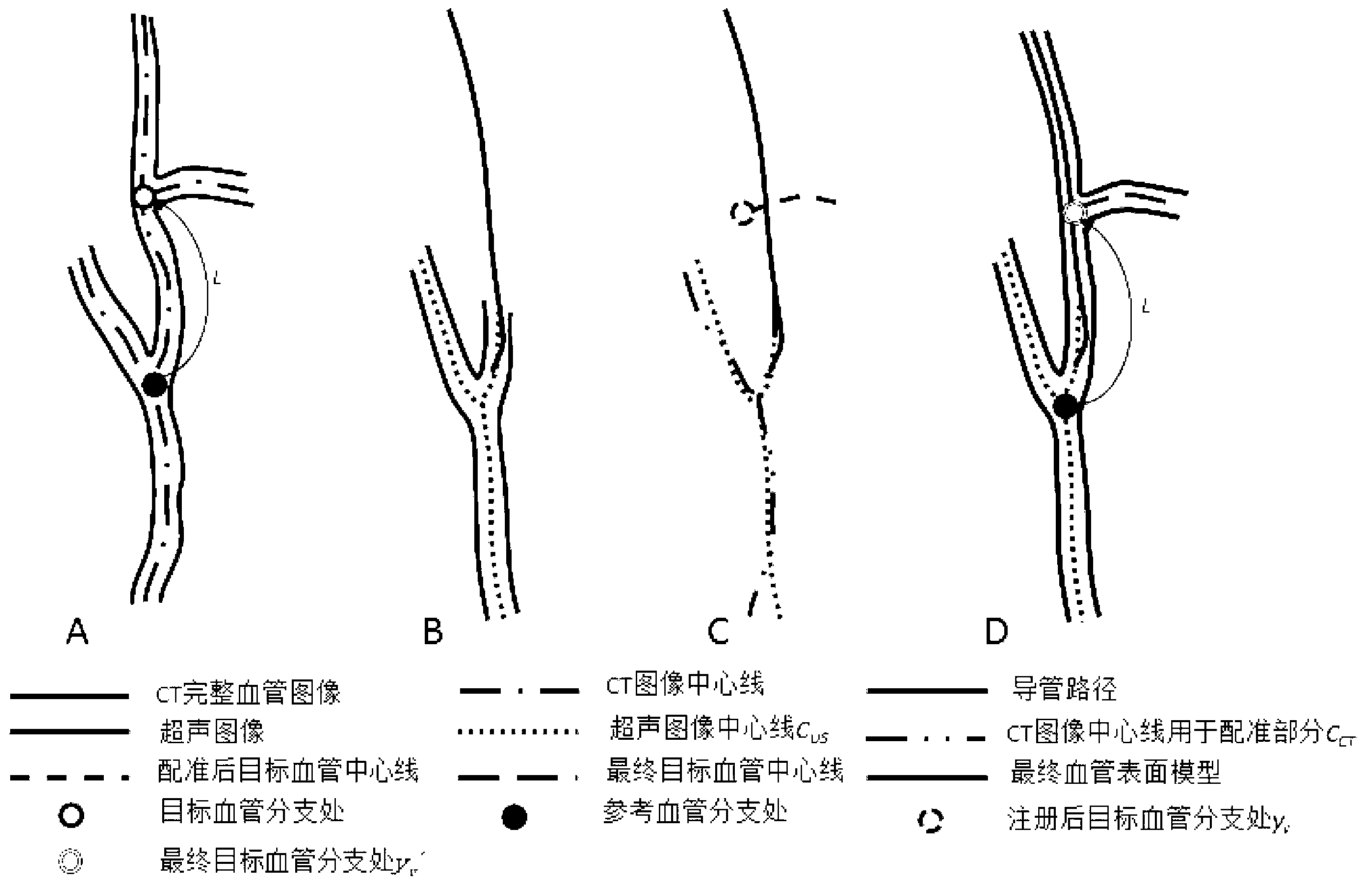
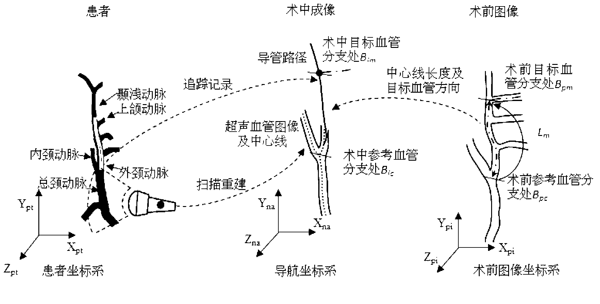
[0025] The present invention studies the problem that ultrasound cannot obtain images of blood vessels under bones, and designs a set of blood vessel visualization devices and methods based on catheter paths to expand ultrasound imaging areas.

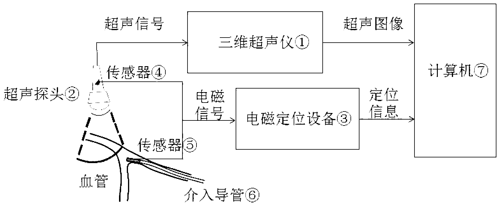
[0026] Vascular minimally invasive interventional surgery can effectively treat diseases under minimally invasive conditions. Three-dimensional Doppler ultrasound is a safe and real-time intraoperative imaging method to obtain vascular morphology, which can accurately and quickly provide intraoperative vascular images to guide the catheter to the target position. However, ultrasound cannot penetrate strong reflectors such as bones to obtain images of blood vessels beneath them for catheter navigation. In the present invention, the catheter path is used to replace the spatial position and direction of the blood vessel in the ultraso...


PUM


Abstract

The invention belongs to the technical field of surgical navigation, and particularly relates to an extension ultrasound vascular imaging device and method based on a catheter path. The extension ultrasound vascular imaging device comprises a three-dimensional ultrasound instrument, a set of electromagnetic positioning equipment, an interventional catheter and a computer, wherein the three-dimensional ultrasound instrument scans a vessel through an ultrasound probe to obtain a three-dimensional image. The electromagnetic positioning equipment comprises a magnetic field generator and two positioning sensors with six degrees of freedom, wherein the magnetic field generator defines a space coordinate system, one positioning sensor with six degrees of freedom is installed on the ultrasound probe to obtain the space coordinate of the ultrasound image, and the other positioning sensor with six degrees of freedom is embedded in an inflexible portion at the front end of the catheter to obtain the space coordinate of the front end of the catheter. The three-dimensional ultrasound instrument and the electromagnetic positioning equipment are both connected with the computer, and the obtained three-dimensional ultrasound image and the obtained catheter path are rebuilt in the same coordinate system through the computer. The extension ultrasound vascular imaging device can position branches of the vessel after the vessel of an area deforms, wherein ultrasound imaging cannot be conducted on the area.

Description

technical field [0001] The invention belongs to the technical field of surgical navigation, and in particular relates to an extended ultrasonic angiography method and device based on a catheter path. Background technique [0002] Vascular minimally invasive interventional surgery is guided by medical imaging equipment, with the help of a catheter along the lumen of the blood vessel to reach a distant lesion (such as coronary artery, carotid artery or tumor blood vessel), and then perform minimally invasive treatment on the lesion An emerging medical procedure. During the operation, the catheter is inserted into the blood vessel of the human body with the cooperation of the guide wire. Generally, a certain part of the human body is punctured first, and then the distal end of the catheter is sent into a specific treatment position along the guide wire, and then the guide wire is pulled out to complete the corresponding treatment process. Vascular minimally invasive intervent...

Claims


Application Information

Patent Timeline
Patent Type & Authority Applications(China)
IPC IPC(8): A61B8/12A61B8/08
Inventor 栾宽李金梁洪王磊张立伟郭良
Owner HARBIN ENG UNIV
Why Eureka
Try Eureka