
Patsnap Eureka
For R&D, Patsnap Eureka makes reading and utilizing patents & technical documents easy.
Patsnap Eureka AIR
Designed for self-driven R&D workflows. Generate viable solutions, solve complex R&D challenges, empower your innovation with AI.

Patsnap Eureka Materials
Designed for material experts only. Revolutionize your material R&D, from search, analyze, to developing new materials.

TechResearch
Generate reliable direction feasibility study reports for your R&D in just a few steps.

TechSeek
Discover and master advanced knowledge NOW. Basics, ideas, possibilities, all at once.

TechMind
As an expert in R&D Theories, TechMind can generates customized viable solutions instantly.

TechRisk
Analyze your overall solution with one click, know your potential R&D risks in advance.

TechMonitor
Get weekly tech updates, stay abreast of the latest tech innovations and key insights.
Meibomian gland infrared imaging device
A technology of infrared imaging and meibomian glands, applied in medical science, equipment for testing eyes, diagnosis, etc., can solve problems such as inspection and diagnosis troubles, and achieve the effect of treatment and prevention
- Summary
- Abstract
- Description
- Claims
- Application Information
AI Technical Summary
Problems solved by technology
Method used
Image

Examples
Embodiment 1
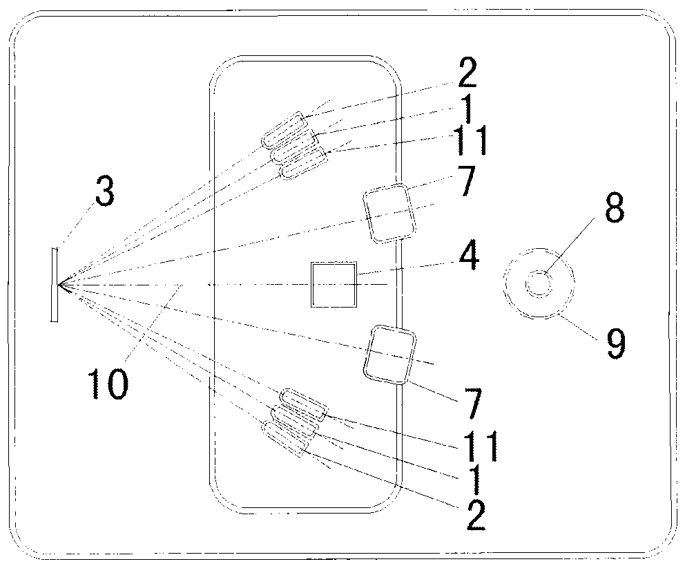
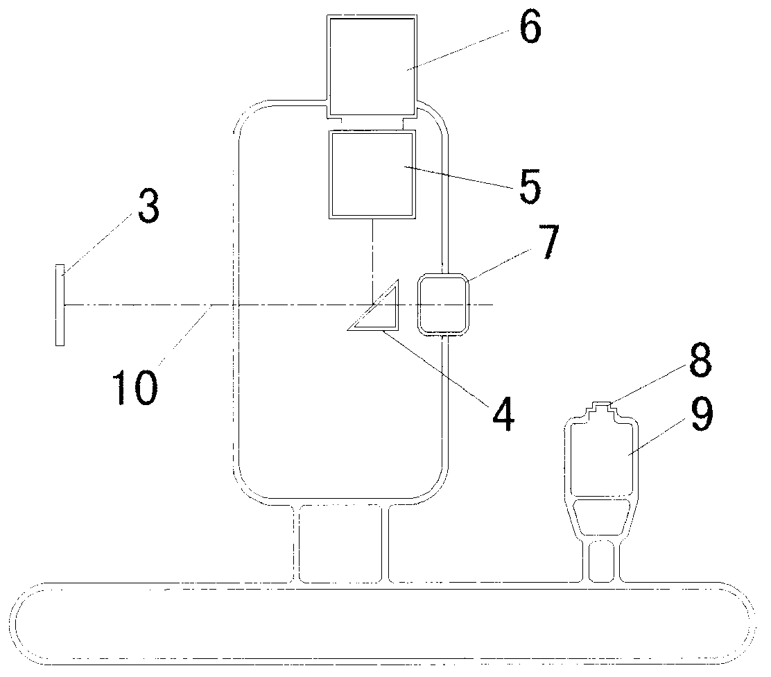
[0022] Embodiment one: if figure 1 , figure 2 As shown, a meibomian gland infrared imaging device, its device main body includes an infrared lighting device 1, a visible light lighting device 2, a focusing light device 11, a coated mirror 4, a focusing lens 5, an infrared camera 6, and a microscope observation device 7 .
[0023] The infrared illuminating device 1 includes two or more sets of infrared point light sources (infrared light beams), and each infrared light point light source (infrared light beams) is symmetrically distributed according to the main optical axis 10 of meibomian glands reflecting infrared light. In this embodiment, two beams of infrared light are used as the infrared illuminating device 1 to act as the main illuminating device. The infrared light emitted by the infrared illuminating device 1 is irradiated on the meibomian glands of the subject, and the reflected infrared light on the meibomian glands Reflected by the coated mirror 4, it enters the ...
Embodiment 2
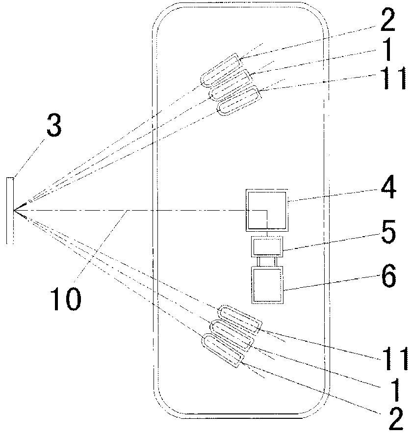
[0033] Such as image 3 As shown, this embodiment is a portable solution of the present invention. The difference between this embodiment and Embodiment 1 is that this embodiment is small and portable, and the microscope observation device 7 is omitted, and front and rear, left and right, up and down and other mechanical movement devices. And the desktop device of embodiment one has microscope observation device 7 and mechanical movement devices such as front and rear, left and right, up and down.
[0034] In addition, this embodiment considers from the perspective of compactness and portability, and the data exchange between the main part of the device and the computer is carried out in a wireless manner. However, the data exchange between the desktop device and the computer in Embodiment 1 is carried out in a cable mode (wireless mode can also be used).
Embodiment 3
[0036] The difference between this embodiment and Embodiment 1 is that the microscope observation device 7 is a single-lens observation device, which is located at the rear end of the coated mirror 4 , and visible light enters the microscope observation device 7 through the coated mirror 4 .
[0037] The coated reflector 4 is a reflector with low reflectivity to visible light. The coated mirror 4 is a 45-degree mirror, which turns 90 degrees to reflect the incident infrared light, and then enters the infrared camera 6 through the focusing lens 5; the incident visible light does not change the optical path transmission, and then enters the microscope observation device 7.
PUM
| Property | Measurement | Unit |
|---|---|---|
| Wavelength | aaaaa | aaaaa |
Abstract
Description
Claims
Application Information
 - R&D Engineer
- R&D Manager
- IP Professional
- Industry Leading Data Capabilities
- Powerful AI technology
- Patent DNA Extraction
Browse by: Latest US Patents, China's latest patents, Technical Efficacy Thesaurus, Application Domain, Technology Topic, Popular Technical Reports.
© 2024 PatSnap. All rights reserved.Legal|Privacy policy|Modern Slavery Act Transparency Statement|Sitemap|About US| Contact US: help@patsnap.com