Method for partitioning brainstem areas automatically from MR (magnetic resonance) sequence images

An automatic segmentation and image-based technology, applied in image analysis, image data processing, instruments, etc., can solve the problems of inconsistent segmentation effect standards, low segmentation efficiency, and labor and material resources consumption, and achieve standardized segmentation results. The effect of reducing the degree of intervention

Inactive Publication Date: 2014-01-01
钟映春
View PDF2 Cites 23 Cited by
  • Summary
  • Abstract
  • Description
  • Claims
  • Application Information

AI Technical Summary

Problems solved by technology

In the past, the segmentation of regions of interest such as the brainstem and facial and auditory nerves was done manually, which required a lot of manpower and material resources, and the segmentation efficiency was low, and the segmentation effect standards were different.

Method used


Image

  • Method for partitioning brainstem areas automatically from MR (magnetic resonance) sequence images
  • Method for partitioning brainstem areas automatically from MR (magnetic resonance) sequence images
  • Method for partitioning brainstem areas automatically from MR (magnetic resonance) sequence images

Examples


Embodiment Construction

[0020] The present invention will be further elaborated below in conjunction with the accompanying drawings.

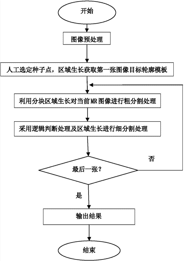
[0021] Such as figure 1 As shown, the specific implementation process of the method for automatically segmenting brainstem regions from sequence MR images of the present invention is as follows:

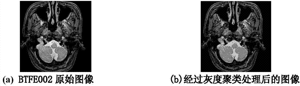
[0022] 1. Neural sequence MR image preprocessing

[0023] The preprocessing is mainly to carry out gray-level clustering processing on all neural sequence MR images, using the method of fuzzy K-means clustering, K is a natural number, clustering the gray-level clustering of all MR images to be processed into 5-level gray-scale images, respectively Represents background, highlighted connective tissue and eyeballs, bones, nervous tissue such as the cerebellum and brainstem, and other tissues.

[0024] The principle of gray-level clustering processing for MR images is: fuzzy cluster analysis is one of the main techniques of unsupervised pattern recognition, which mainly seeks...


PUM


Abstract

The invention relates to a method for partitioning brainstem areas automatically from MR (magnetic resonance) sequence images. The method is used for partitioning the brainstem areas automatically and continuously from the human body encephalic MR sequence images, so as to offer technological bases for reconstruction of 3D model of the encephalic tissues to doctors to judge the state of an illness of the patient efficiently and intuitively. The method comprises steps of (1) preprocessing: carrying out gray level clustering analysis on all MR nerve sequence images, and clustering the gray levels of all the MR images to be processed to be five-level gray level images; (2) selecting seed point pixel, obtaining the outline border of the brainstem area of the first MR image through a neighbor region growing algorithm based on the image area growing rate; (3) roughly partitioning the current MR image through a block region growing method; (4) repartitioning the roughly partitioned MR images through logical judgment treatment and the region growing method; and (5) repeating the step (3) and the step (4) till finishing partitioning all MR images.

Description

technical field [0001] The invention is a method applicable to the automatic segmentation of specific targets in MR sequence images in medicine, and belongs to the technical field of medical images, in particular, it relates to a method for realizing continuous Automatic segmentation, to provide the technical basis for doctors to reconstruct the 3D model of intracranial tissue, so as to judge the patient's condition more efficiently and intuitively. The method of automatically segmenting the brainstem region from the sequence of MR images. Background technique [0002] Image segmentation refers to the technology and process of dividing an image into regions with different characteristics and extracting objects of interest. Image segmentation has been widely used in practice, such as computer vision, remote sensing and biomedical image analysis, as well as military, sports, agricultural engineering and other fields. In a nutshell, it is only necessary to extract and measure ...

Claims


Application Information

Patent Timeline
Patent Type & Authority Applications(China)
IPC IPC(8): G06T7/00
Inventor 钟映春赖丹练张勇黄英罗唯师钱东翔
Owner 钟映春
Why Eureka
Try Eureka