Automatic blood cell recognition device and operating method thereof

A technology for identifying devices and blood cells, which is used in measurement devices, particle and sedimentation analysis, individual particle analysis, etc., and can solve the problems of difficulty in improving identification accuracy, poor staining, and complex automatic analysis algorithms for blood cells.

Active Publication Date: 2014-02-26
SHANDONG UNIV
View PDF4 Cites 20 Cited by
  • Summary
  • Abstract
  • Description
  • Claims
  • Application Information

AI Technical Summary

Problems solved by technology

In the existing automatic diagnosis and analysis of blood cells based on microscopic images, the two-dimensional color or grayscale images of blood smears taken under the microscope are basically the research objects, because these images can only provide limited information such as grayscale or color. information, which makes the automatic analysis algorithm of blood cells relatively complex, and can only analyze blood cells from the perspective of shape and color, and cannot conduct a more in-depth analysis of diseased cells from the perspective of biochemistry
Moreover, due to the complex shape of the cells in the image, the cells often overlap, and the poor staining of the specimen, it brings many difficulties to the automatic image recognition of blood cells based on traditional imaging technology. There are many types of blood cells and subtle morphological differences, which make the recognition accuracy difficult. bottleneck of improvement

Method used


Image

  • Automatic blood cell recognition device and operating method thereof
  • Automatic blood cell recognition device and operating method thereof
  • Automatic blood cell recognition device and operating method thereof

Examples


Embodiment 1

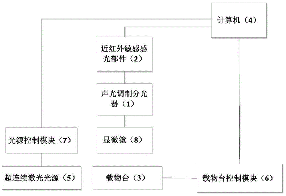
[0048] like figure 1 shown.

[0049] An automatic blood cell identification device, comprising a laser light source part, a microscope collection part and a stage part respectively connected to a computer;

[0050] The laser light source part includes a light source control module and a supercontinuum laser light source;

[0051] The microscope acquisition unit includes a near-infrared sensitive photosensitive component, an acousto-optic modulation beam splitter, and a microscope connected in sequence with a computer; under supercontinuum laser irradiation, blood cell slices enter the microscope beam splitter through the objective lens and eyepiece optical path of the microscope, and then The infrared sensitive component realizes imaging, wherein the beam splitter is used to decompose the mixed light into simple light of each band;

[0052] The stage part includes a stage control module and a stage, and the stage control module communicates with a computer through an RS-232 ...

Embodiment 2

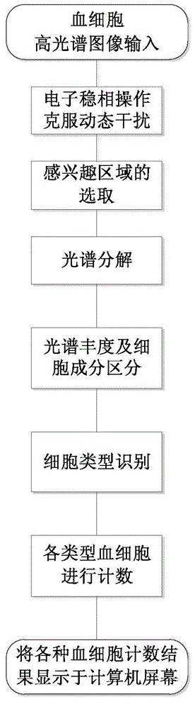
[0057] like figure 2 shown.

[0058] A working method of the automatic blood cell identification device as described in embodiment 1, comprising the following steps:

[0059] (1) The hyperspectral image of blood cells acquired by the near-infrared sensitive photosensitive component through a microscope is sent to the computer through a transmission link; the transmission link is Ethernet;

[0060] (2) In order to overcome the dynamic interference, the hyperspectral image obtained by computer is used for electronic phase stabilization operation, the specific process is as follows:

[0061] Step1. Each image is divided into macroblocks, preferably, the size of the macroblocks is 16×16 pixels;

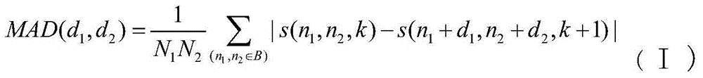
[0062] Step2. Taking the first image as the reference frame, for each macroblock in the first image, in the corresponding search area in each subsequent image, search for the best matching block according to the matching criterion, that is, according to The matching with the smallest ...


PUM


Abstract

The invention provides an automatic blood cell recognition device which comprises a laser light source part, a microscope acquisition part and an objective table part which are respectively connected with a computer. The blood cells can be automatically recognized without staining or utilizing an antibody/antigen reaction on the cell surface, and the blood cell recognition is realized by utilizing cell morphology and spectral characteristic change according to a hyperspectral image. The system is a human blood cell recognition technology with the technical characteristic of automatic non-destructiveness.

Description

technical field [0001] The invention relates to an automatic blood cell identification device and a working method, belonging to the technical field of blood cell identification. Background technique [0002] After nearly 50 years of development, computer-aided blood cell analysis technology is not limited to routine blood cell analysis. In recent years, it has added many expanded functions, such as counting and analyzing reticulocytes (RET) (Bayer ADVIA120; Sysmex SE-9000 / SE-9500 / XE-2100 type; CoulterMAXM / STKS / HmX / GEN.S type; Abbott CELL-DYN3200 / 3500R / 4000 type, etc.), immature cell analysis and nucleated red blood cell analysis function (Sysmex SF-3000 / SE-9000 / SE-9500 / XE-2100; Bayer ADVIA120; Abbott CELL-DYN3500 / 4000; Coulter HmX / LH750 / GEN.SSystem2, etc.), and some instruments combine some functions of the flow cytometer To the blood cell analyzer, so that the analysis results of certain lymphocyte subsets can be obtained during routine blood cell analysis (for example, ...

Claims


Application Information

Patent Timeline
Patent Type & Authority Applications(China)
IPC IPC(8): G01N15/10
Inventor 郑成云刘治李晓梅陈颖洁
Owner SHANDONG UNIV
Why Eureka
Try Eureka