Wound imaging device

An imaging device and wound technology, applied in medical science, sensors, diagnostic recording/measurement, etc., can solve the problems of poor accuracy, slow measurement speed, and risk of infection, and achieve the effect of high precision and fast measurement speed

Inactive Publication Date: 2014-03-19
QINGDAO ANXIN MEDICAL DEVICES
View PDF5 Cites 0 Cited by
  • Summary
  • Abstract
  • Description
  • Claims
  • Application Information

AI Technical Summary

Problems solved by technology

[0002] The degree of wound healing can be reflected by the size of the wound. In the prior art, the size of the wound is usually measured by the medical staff using a ruler and calculated manually. This not only has the risk of infection, but also the measurement speed is slow and the accuracy is poor.

Method used


Image

  • Wound imaging device

Examples


Embodiment 1

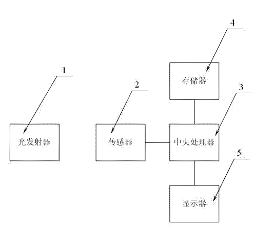
[0014] Example 1, such as figure 1 As shown, this embodiment discloses a wound imaging device, which includes a light emitter 1, the light emitted by the light emitter is reflected by the wound, and then received by the sensor 2, and the central processing unit 3 processes the signal received by the sensor. After encoding and imaging, the imaging results are stored in the memory 4 and displayed on the display 5 as output. The wavelength of the light emitted by the light emitter is 800nm-900nm. The light emitter irradiates the wound surface at an angle of 30°-50°. The light emitter is an LED bulb. The sensor is a digital monochrome photoreceptor.

[0015] The wound imaging device disclosed in this embodiment does not directly contact the patient's wound, has no risk of infection, has fast measurement speed and high precision, and is suitable for popularization and application in clinical practice.


PUM


Abstract

The invention discloses a wound imaging device which comprises an optical transmitting set. Light emitted by the optical transmitting set is reflected by a wound and then is received by a sensor, a central processing unit carries out coded imaging on the signals received by the sensor, and imaging results are stored in a memorizer and are displayed and output through a displayer. The wound imaging device does not make contact with the wound directly, infection risks do not exist, measuring speed is fast, precision is high, and the wound imaging device is suitable for being popularized and applied in clinical practice.

Description

technical field [0001] The invention relates to an imaging device, in particular to a wound imaging device. Background technique [0002] The degree of wound healing can be reflected by the size of the wound. In the prior art, the size of the wound is usually measured by medical personnel using a ruler and calculated manually. This not only has the risk of infection, but also has slow measurement speed and poor accuracy. Contents of the invention [0003] The technical problem to be solved by the present invention is to provide a wound imaging device that can quickly and accurately obtain and calculate the size of the wound. [0004] In order to solve the above technical problems, the present invention adopts the following technical solutions: [0005] A wound imaging device, the device includes a light emitter, the light emitted by the light emitter is reflected by the wound, and then received by the sensor, and the central processing unit performs encoding and imaging o...

Claims


Application Information

Patent Timeline
Patent Type & Authority Applications(China)
IPC IPC(8): A61B5/00
Inventor 不公告发明人
Owner QINGDAO ANXIN MEDICAL DEVICES
Why Eureka
Try Eureka