Magnetic resonance parameter imaging method and system

An imaging method and magnetic resonance image technology, applied in medical science, sensors, diagnostic recording/measurement, etc., can solve the problems of slow imaging speed of magnetic resonance parameters, affecting the quality of parameter imaging, and easy introduction of motion artifacts, etc.

Active Publication Date: 2014-04-09
SHENZHEN INST OF ADVANCED TECH
View PDF3 Cites 22 Cited by
  • Summary
  • Abstract
  • Description
  • Claims
  • Application Information

AI Technical Summary

Problems solved by technology

[0003] However, due to the need to continuously sample a series of images, the speed of MRI parametric imaging is relatively slow, which can easily introduce motion artifacts and affect the quality of parametric imaging

Method used


Image

  • Magnetic resonance parameter imaging method and system
  • Magnetic resonance parameter imaging method and system
  • Magnetic resonance parameter imaging method and system

Examples


Embodiment Construction

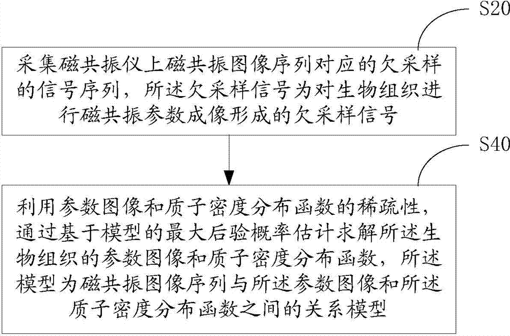
[0069] Such as figure 1 Shown, in one embodiment, a kind of magnetic resonance parametric imaging method, comprises the following steps:

[0070] Step S20 , acquiring an undersampled signal corresponding to the magnetic resonance image sequence on the magnetic resonance apparatus, where the undersampled signal is an undersampled signal formed by performing magnetic resonance parameter imaging on the biological tissue.

[0071] Step S40, using the sparsity of the parameter image and the proton density distribution function, solving the parameter image and the proton density distribution function of the biological tissue through the maximum a posteriori probability estimation based on the model, the model is the magnetic resonance image sequence and the parameter Model the relationship between the image and the proton density distribution function.

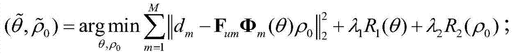
[0072] In one of the embodiments, the undersampled signal d corresponding to the mth frame image of the magnetic resonance image ...


PUM


Abstract

A magnetic resonance parameter imaging method includes the steps of acquiring an under-sampling signal corresponding to a magnetic resonance image sequence on a magnetic resonance imager; estimating and solving a parameter image and proton density distribution function of biological issue through sparsity in parameter image and proton density distribution functions and model-based maximum posterior probability. The under-sampling signal is formed by application of magnetic resonance parameter imaging on the biological issue. A model for maximum posterior probability is a model of relation between the magnetic resonance image sequence and the parameter image and proton density distribution function. The method has the advantages that the parameter image and proton density distribution function of the biological tissue is directly estimated through the under-sampling signal and error propagation caused by the application of an under-sampling rebuilt magnetic resonance image in estimating biological tissue parameters is avoided. The sparsity in the parameter image and proton density distribution function is introduced, and image artifacts caused by under-sampling can be effectively suppressed. In addition, the invention further provides a magnetic resonance parameter imaging system.

Description

【Technical field】 [0001] The invention relates to the field of magnetic resonance imaging, in particular to a magnetic resonance parameter imaging method and system. 【Background technique】 [0002] Magnetic Resonance Imaging (MRI) is an advanced non-invasive medical diagnostic tool. It uses the principle of nuclear magnetic resonance (NMR) to draw information about the internal anatomical structure and physiological functions of biological tissues. Among them, magnetic resonance parametric imaging (such as T1, T2, T2*, Diffusion Tensor, etc.) provides a method that can quantitatively analyze the biochemical properties of living tissues. [0003] However, due to the need to continuously sample a series of images, the speed of MRI parametric imaging is relatively slow, which easily introduces motion artifacts and affects the quality of parametric imaging. Accelerated MRI parametric imaging is also an important and difficult issue in its clinical application. 【Content of in...

Claims


Application Information

Patent Timeline
Patent Type & Authority Applications(China)
IPC IPC(8): A61B5/055
Inventor 彭玺梁栋刘新郑海荣
Owner SHENZHEN INST OF ADVANCED TECH
Why Eureka
Try Eureka