Position information based ultrasonic wide view imaging method

A position information and wide-view imaging technology, which is applied in the field of medical ultrasound wide-view imaging, can solve the problems of large amount of calculation, EFOV technology cannot meet the real-time requirements, and it is difficult to achieve real-time, so as to reduce the amount of calculation and solve image registration Time-consuming, simple effects to implement

Inactive Publication Date: 2014-04-30
SOUTH CHINA UNIV OF TECH
View PDF5 Cites 9 Cited by
  • Summary
  • Abstract
  • Description
  • Claims
  • Application Information

AI Technical Summary

Problems solved by technology

Wide-field imaging technology mainly includes three parts: image acquisition, image registration, and image mosaic; among them, image registration is the core of wide-field imaging. In order to accurately obtain the relative displacement and deflection between two adjacent images, image registration It is nec

Method used


Image

  • Position information based ultrasonic wide view imaging method
  • Position information based ultrasonic wide view imaging method
  • Position information based ultrasonic wide view imaging method

Examples


Embodiment 1

[0027] The present invention will be described in further detail below in conjunction with the embodiments and accompanying drawings, but the embodiments of the present invention are not limited thereto.

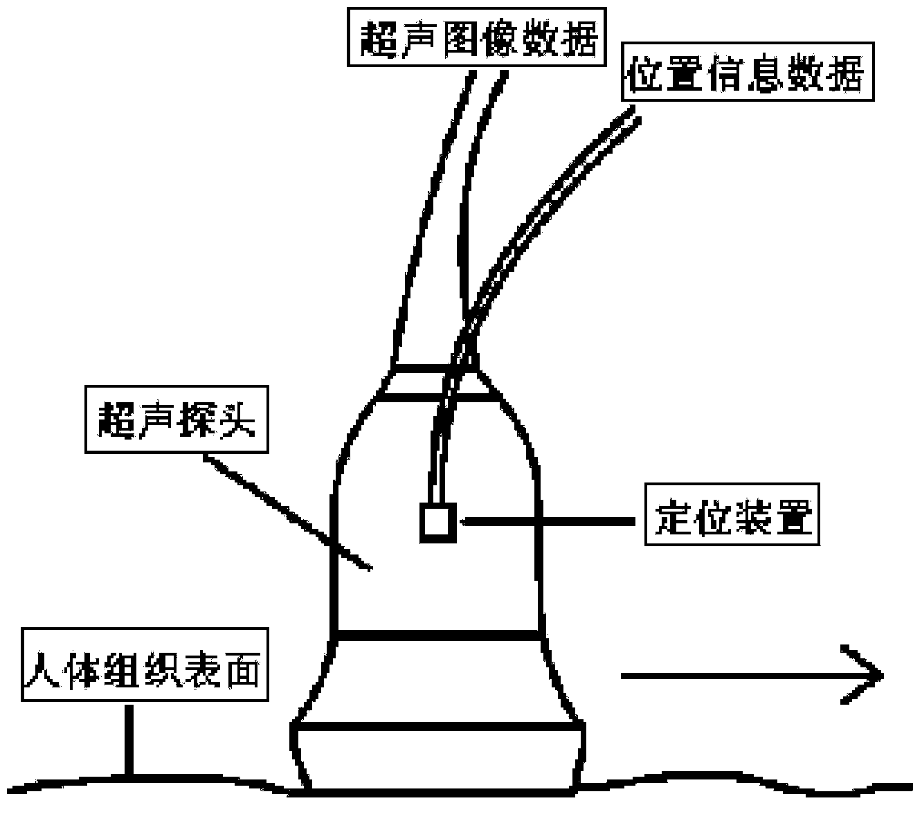
[0028] The ultrasonic wide-view imaging method of this embodiment is realized by adopting an ultrasonic probe and installing a positioning device on the ultrasonic probe. The positioning device of this embodiment uses the miniBIRD electromagnetic positioning system developed by Ascension Company, which includes the following steps:

[0029] 1) if figure 1 As shown, when collecting images on the surface of human tissue, the ultrasonic probe moves laterally along a straight line or an approximate straight line (as shown by the arrow in the figure), the system obtains the ultrasonic two-dimensional image sequence during the scanning process, and obtains the image through the positioning device The position information of the acquired ultrasonic two-dimensional image sequence su...

Embodiment 2

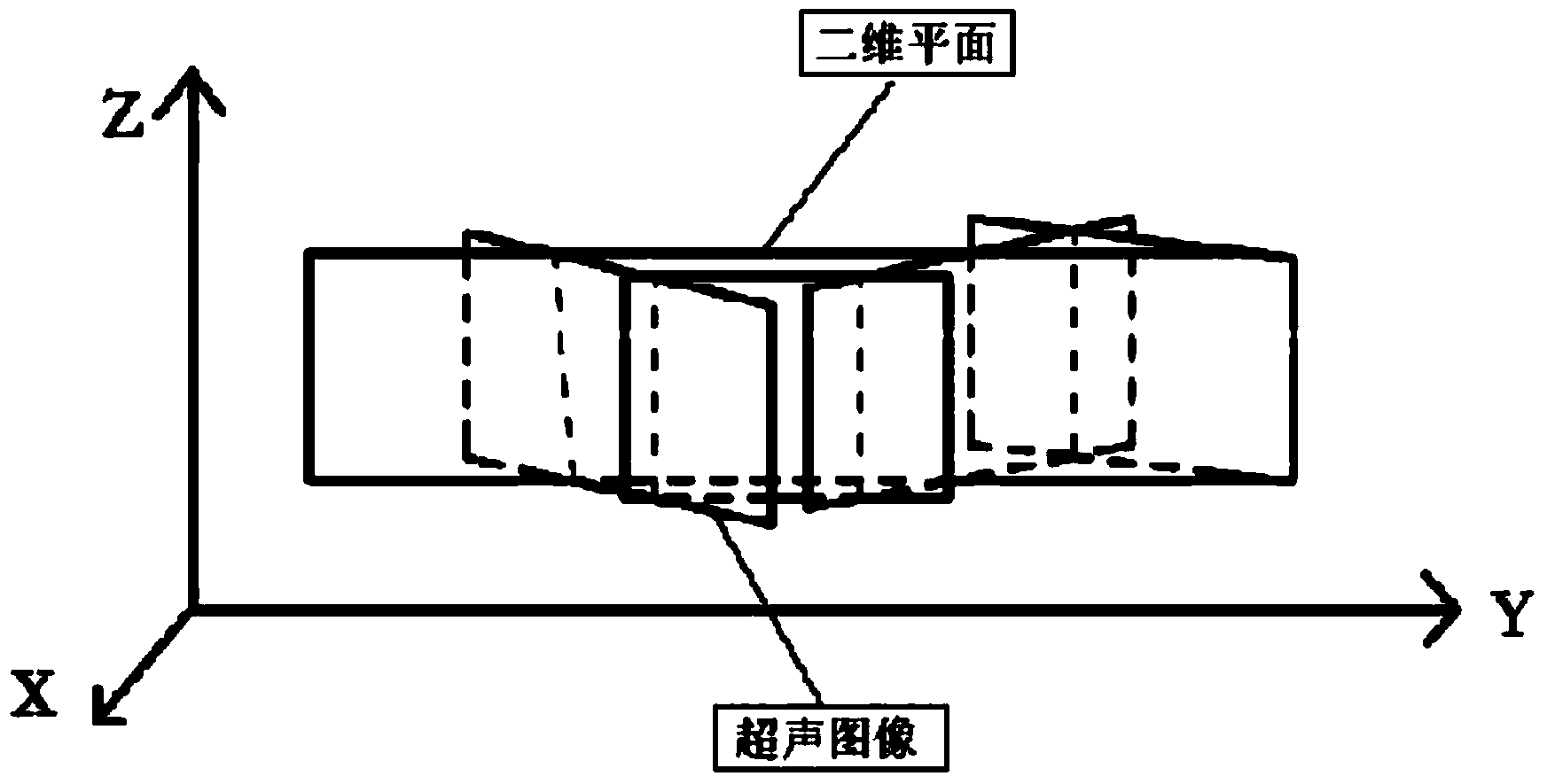
[0036] The main features of this embodiment are: in step 4), each frame image is mapped to a two-dimensional plane according to its spatial position information in the world coordinate system, and the selection of this two-dimensional plane is to place all frame images on the plane Linear fitting is performed to obtain a two-dimensional plane, so that the average distance between each frame image and the two-dimensional plane is the shortest, and a panoramic image closer to the actual image is obtained. All the other are with embodiment 1.


PUM


Abstract

The invention discloses a position information based ultrasonic wide view imaging method. The method includes the following steps that (1) when acquiring images, an ultrasonic probe moves along a straight line or an approximate straight line, a system acquires an ultrasonic two-dimensional image sequence in the scanning process, and position information of the images is obtained through a positioning device; (2) time is set for the system, each frame image acquired is corresponding to the position information thereof; (3) spatial calibration is performed to enable each frame image to be transformed into a coordinate system of the positioning device, position and direction information acquired by the positioning device in the moving process of the ultrasonic probe is recorded in a world coordinate system, and coordinates of each frame image in the world coordinate system are obtained by final transformation for the convenience of display; (4) each frame image is mapped onto a preset two-dimensional plane according to the spatial position information thereof in the world coordinate system to form a wide view image. By the method, calculated amount of wide view imaging can be reduced, and real-time performance can be improved.

Description

technical field [0001] The invention relates to a medical ultrasonic wide-view imaging method, in particular to an ultrasonic wide-view imaging method based on position information, and belongs to the technical field of medical ultrasonic wide-view imaging. Background technique [0002] Medical ultrasound imaging is one of the important diagnostic methods in modern clinical medicine. Due to the limited divergence angle of the ultrasound beam emitted by the traditional ultrasound probe and the size of the probe, it can only generate ultrasound images with a narrow field of view and cannot reflect The overall image of the detected area affects the accuracy and speed of medical diagnosis to a certain extent. [0003] In response to this problem, in 1996, Weng et al. proposed Extended field-of-view ultrasound, which collects a series of ultrasound images and uses image registration technology to stitch them together in real time to form an image with a larger observation field o...

Claims


Application Information

Patent Timeline
IPC IPC(8): A61B8/00G06T7/00
Inventor 黄庆华赵夏
Owner SOUTH CHINA UNIV OF TECH
Why Eureka
Try Eureka