Feature subspace integration method for biological cell microscope image classification

A technology of characteristic subspace and biological cells, which is applied in the direction of instruments, character and pattern recognition, computer components, etc., can solve the problems of poor image classification effect and poor classification accuracy

Active Publication Date: 2014-07-02
XIAN JIAOTONG LIVERPOOL UNIV
View PDF4 Cites 3 Cited by
  • Summary
  • Abstract
  • Description
  • Claims
  • Application Information

AI Technical Summary

Problems solved by technology

[0006] The purpose of the present invention is to provide a feature subspace integration method for biological cell microscope ima

Method used


Image

  • Feature subspace integration method for biological cell microscope image classification
  • Feature subspace integration method for biological cell microscope image classification
  • Feature subspace integration method for biological cell microscope image classification

Examples


Embodiment

[0050] The feature subspace integration method used in this embodiment for biological cell microscope image classification includes the following steps:

[0051] (1) Extract the features of the microscope images of biological cells to be classified by the following methods:

[0052] i) Transform the microscope image of biological cells into different frequency sub-bands (Sub-band), and then perform feature statistics on each frequency sub-band; and

[0053] ii) Using multiple statistical features of the gray level co-occurrence matrix to obtain the global texture features of the microscope image of biological cells; and

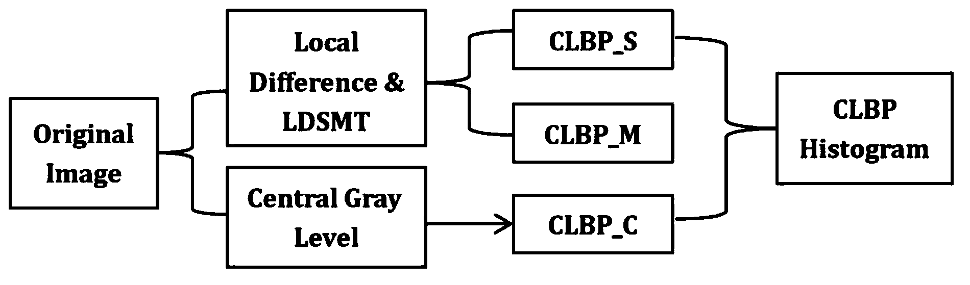
[0054] iii) Extracting local texture features of biological microscope images through full local binary mode;

[0055] (2) Using KPCA to construct feature subspaces for the three image features of the extracted biological cell microscope images, so that each type of biological cell microscope images has three feature subspaces;

[0056] (3) Use the three tr...


PUM


Abstract

The invention discloses a feature subspace integration method for biological cell microscope image classification. The method comprises the following steps: features of biological cell microscope images to be classified are extracted; a feature subspace model is constructed for three extracted image features of the biological cell microscope images by using the kernel principal component analysis (KPCA) to make each type of biological cell microscope images has three feature subspaces; three image features are reconstructed for the biological cell microscope images to be classified by adopting each type of three trained feature subspaces to obtain the reconstruction result of each feature subspace in each type on the image features to be classified, and the classification confidence of the classified images on each type is obtained through the comparison between the reconstruction result of each feature subspace and the originally-extracted image feature vectors; and the images to be classified are classified into the type having the highest confidence. According to the method, the feature dimension can be effectively reduced, the diversity of integration classifiers can be improved, and the classification effect can be further improved.

Description

technical field [0001] The invention belongs to the field of intelligent image analysis, in particular to the classification research of biological cell microscope images, and specifically relates to a feature subspace integration method for biological cell microscope image classification. Background technique [0002] Cell microscopic imaging technology is crucial to understanding the function and structure of cells. For the diagnosis and classification of many diseases, cell biopsy has become the gold standard for diagnosis. However, a large number of pathological biopsy imaging will cause a huge workload for medical image analysts; manual detection may sometimes cause false detections and missed detections due to various external factors (such as fatigue, lack of experience, etc.). Therefore, automatic Cell microscope image detection technology has become a research hotspot in recent years. [0003] A widely used method for detecting the location of subcellular proteins ...

Claims


Application Information

Patent Timeline
IPC IPC(8): G06K9/46G06K9/66
Inventor 张百灵张云港
Owner XIAN JIAOTONG LIVERPOOL UNIV
Why Eureka
Try Eureka