

Computed tomography system with CT respiratory gating
A technology of tomography and respiratory gating, applied in computerized tomography scanners, echo tomography, etc., can solve problems affecting diagnosis and judgment, patient discomfort, difficult operation, etc., to achieve convenient operation, reduce discomfort, and reduce discomfort Sensitive effect
Inactive Publication Date: 2014-07-23
JIANGSU SINOWAYS (ZHONGHUI) MEDICAL TECH CO LTD
View PDF5 Cites 2 Cited by
- Summary
- Abstract
- Description
- Claims
- Application Information
AI Technical Summary
Problems solved by technology
[0004] 1. The detection electrode needs to be installed on the detected part (chest), which brings discomfort to the patient
[0005] 2. The introduction of metal substances will lead to unnecessary metal artifacts, which will affect the diagnosis and judgment
[0006] 3. Use cables and fixing straps to fix the load unit on the chest of the examinee, which is easy to interfere with the chest scanning part during use, and is not easy to operate
Method used
View moreImage
Smart ImageViewing Examples

Examples
Embodiment Construction
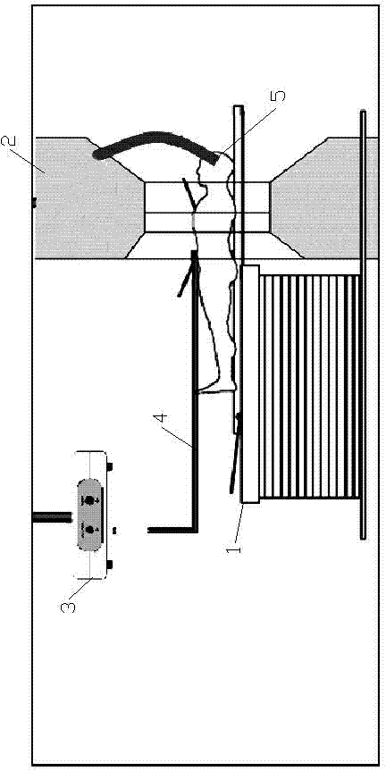
[0016] Such as figure 1 As shown, the present invention includes a bed body 1, a CT barrel frame 2, a sensor terminal 3, a load unit 4 and a detection probe 5 for inner ear pressure. The signal output end of the inner ear pressure detection probe 5 is connected to the signal input end of the CT barrel holder 2 .
[0017] During detection, the signal acquisition end of the inner ear pressure detection probe 5 is placed in the subject's ear, and the movement position of the chest is accurately detected according to the relationship between the ear pressure and the breathing process. Thus, the straight-line CT machine performs respiratory-synchronized scanning in a specific implementation and cycle.
Login to View More PUM
 Abstract
The invention relates to the technical field of computed tomography systems and discloses a CT system with CT respiratory gating. The CT system comprises a bed body, a CT gantry, a sensor terminal, a loading unit and a detection probe for inner ear pressure. The signal output end of the detection probe for inner ear pressure is connected to the input end of the CT gantry. The advanced ear pressure measuring mode is adopted for reflecting respiratory motion and chest motion and combined with CT chest examination to guide control over scanning time and a scanning position. While the CT system plays an effective role in respiratory gating, influences on a patient and image quality are reduced, and the CT system not only is convenient to operate, but also reduces the uncomfortable degree of the examined patient and meanwhile guarantees detection accuracy.
Description
technical field [0001] The present invention relates to the technical field of computer tomography system, that is, CT instrument. Background technique [0002] In order to remove the motion artifacts caused by respiratory motion when the computerized tomography system (CT) scans the chest and heart, it is necessary to use a respiratory monitoring device that can detect chest motion. Multiple scans are taken at the same location where the chest expands. [0003] In order to achieve the purpose of measurement, the current general practice in the industry is to use the chest electrode plate to measure the impedance to reflect the ups and downs of the chest. But this method has some disadvantages in use: [0004] 1. The detection electrode needs to be installed on the detected part (chest), which brings discomfort to the patient. [0005] 2. The introduction of metal substances will lead to unnecessary metal artifacts, which will affect the diagnosis and judgment. [00...
Claims
Login to View More Application Information
Patent Timeline
 IPC IPC(8): A61B6/03
Inventor周宇任敬轶付诗农居小平王涛高飞郜文兵冯海友
OwnerJIANGSU SINOWAYS (ZHONGHUI) MEDICAL TECH CO LTD