Novel retina eye fundus image segmenting method

A fundus image and retina technology, applied in image analysis, image enhancement, image data processing, etc., can solve the problems of wrong blood vessel detection, small blood vessels are easy to lose, and blood vessels are easy to merge

Inactive Publication Date: 2014-09-10
CHONGQING UNIV
View PDF3 Cites 17 Cited by
  • Summary
  • Abstract
  • Description
  • Claims
  • Application Information

AI Technical Summary

Problems solved by technology

[0004] The above-mentioned blood vessel segmentation algorithm still has some defects, including: the segmentation effect of the central reflection area of ​​the blood vessel is poor, and the segmentation ef

Method used


Image

  • Novel retina eye fundus image segmenting method
  • Novel retina eye fundus image segmenting method
  • Novel retina eye fundus image segmenting method

Examples


Embodiment Construction

[0022] The preferred embodiments of the present invention will be described in detail below in conjunction with the accompanying drawings. This embodiment is implemented on the premise of the technical solution of the present invention, and detailed implementation methods and specific operating procedures are provided, but the preferred examples are only for illustrating the present invention , not to limit the protection scope of the present invention.

[0023] The image frames used in this implementation come from a standard database.

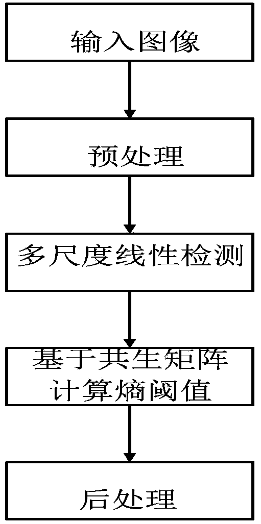
[0024] figure 1 The overall system block diagram of a new retinal fundus image segmentation method provided by the embodiment of the present invention, as shown in the figure: the system block diagram is composed of 4 functional modules: (1) image preprocessing; (2) multi-scale linear detection ; (3) Calculate the optimal entropy threshold based on the gray-gradient co-occurrence matrix; (4) Post-processing

[0025] figure 2 is the prepro...


PUM


Abstract

The invention provides a novel retina eye fundus image segmenting method. The method is characterized in that the best entropy threshold value is calculated by combining multi-scale linear detection and using the gray-level-gradient co-occurrence matrix of an image. Firstly, green components, containing rich blood vessel outline information, in the retina eye fundus image are extracted, and shadow correcting, noise reducing, CLAHE and other preprocessing are performed on the green components; secondly, multi-scale and multi-direction linear detection is performed on blood vessels of the retina eye fundus image according to morphological structure characteristics of the blood vessels, and image responses of different scales are fused to obtain the characteristics of the blood vessels; finally the best entropy threshold value of the image is calculated on the basis of the gray-level-gradient co-occurrence matrix of the image, and segmentation is performed. The method is high in segmenting accuracy, capable of extracting more fine blood vessels, high in calculating speed, very good in robustness and suitable for segmentation of the normal or lesion retina eye fundus image.

Description

technical field [0001] The invention relates to the field of computer image processing, in particular to a retinal fundus image segmentation method based on combining multi-scale linear detection and using a gray-gradient co-occurrence matrix to obtain an optimal entropy threshold, especially suitable for normal retinal fundus image segmentation. Background technique [0002] Retinal blood vessels are an important part of the systemic microcirculatory system, and changes in their morphology are closely related to the severity of cardiovascular diseases such as diabetes, hypertension, cerebrovascular sclerosis and coronary arteriosclerosis. By extracting retinal blood vessels, analyzing their characteristics, such as vessel diameter and curvature, and measuring and analyzing related parameters, cardiovascular diseases can be predicted to a large extent, so as to implement scientific preventive intervention and drug treatment . [0003] There are still many segmentation algor...

Claims


Application Information

Patent Timeline
IPC IPC(8): G06T7/00G06T5/00
Inventor 张思杰翟丽红邓蕊戴阳徐鹏曾孝平
Owner CHONGQING UNIV
Why Eureka
Try Eureka