
A cerebral arterial thrombosis image segmentation method

A technology for ischemic stroke and image segmentation, which is applied in the field of medical image processing and can solve problems such as irregular shape, blurred boundary, and uneven internal brightness.

Active Publication Date: 2019-05-10
江门市中心医院 +1
View PDF17 Cites 2 Cited by
  • Summary
  • Abstract
  • Description
  • Claims
  • Application Information

AI Technical Summary

Problems solved by technology

However, the existence of the "ischemic penumbra" makes the entire cerebral infarction lesion have the characteristics of blurred boundary, irregular shape and uneven internal brightness on DWI images, which brings challenges to accurately segment the location and size of the lesion area

Method used


Image

  • A cerebral arterial thrombosis image segmentation method
  • A cerebral arterial thrombosis image segmentation method
  • A cerebral arterial thrombosis image segmentation method

Examples


Embodiment 1

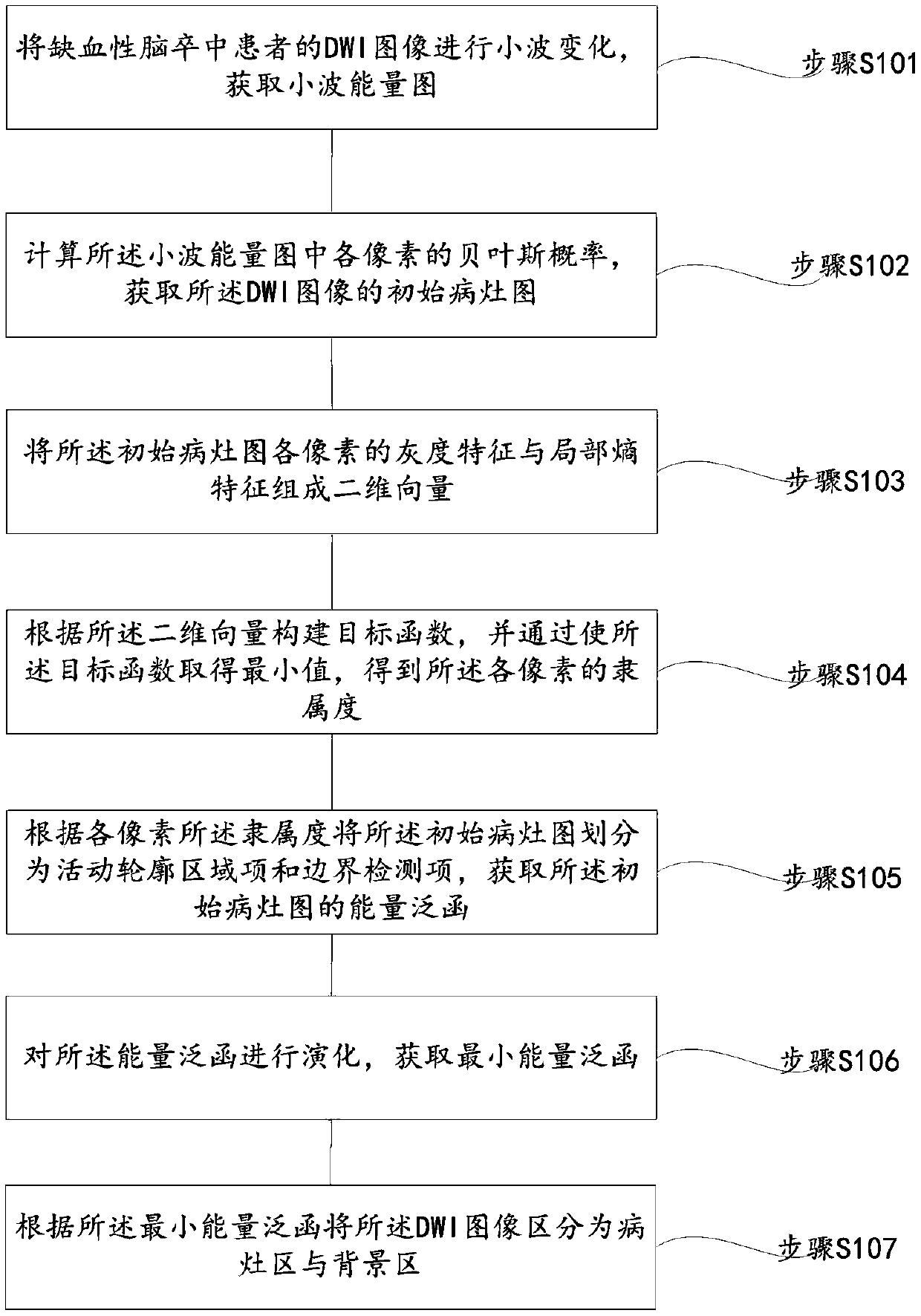
[0065] refer to figure 1 , which shows a flow chart of the steps of an embodiment of ischemic stroke image segmentation in the present invention, which may specifically include the following steps:

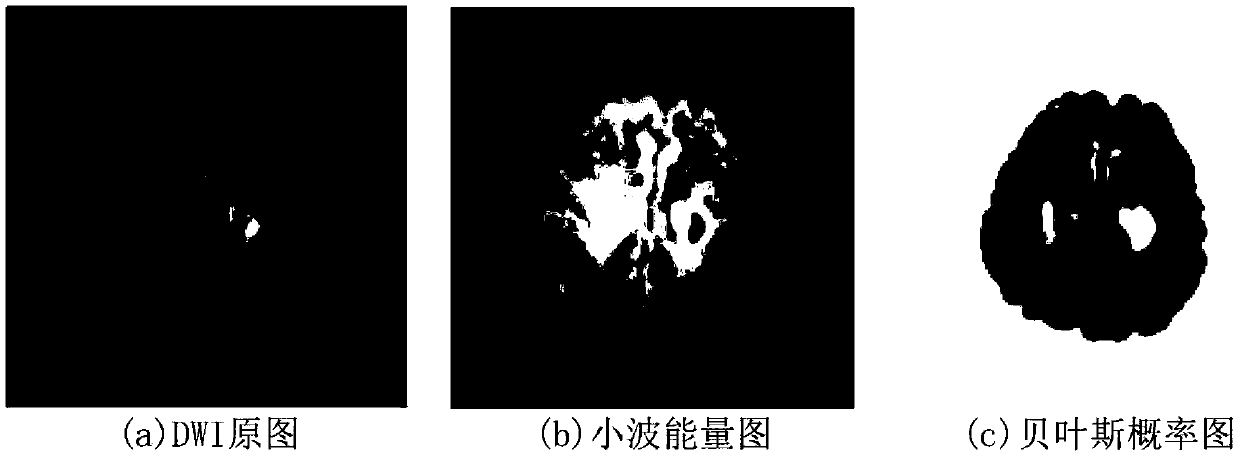
[0066] Step S101, performing wavelet transformation on the DWI image of a patient with ischemic stroke to obtain a wavelet energy map;

[0067] Since the nerve cells in the infarcted area of ​​patients with acute cerebral infarction have been necrotic and cannot metabolize normally, and the surrounding "ischemic penumbra" and edema appear, the water molecules in the "ischemic penumbra" and edema area in the infarcted area The distribution loses its regularity and is significantly different from normal brain tissue. According to the above reasons, the DWI image of the patient with ischemic stroke is analyzed to accurately distinguish the lesion area from the background area.

[0068] The initial position of the model contour curve has a great influence on the segmentation accurac...

Embodiment 2

[0108] refer to Figure 4 , which shows a flow chart of the steps of an embodiment of ischemic stroke image segmentation in the present invention, which may specifically include the following steps:

[0109] Step S201, performing wavelet transformation on the DWI image of the patient with ischemic stroke to obtain a wavelet energy map;

[0110] Since the nerve cells in the infarcted area of ​​patients with acute cerebral infarction have been necrotic and cannot metabolize normally, and the surrounding "ischemic penumbra" and edema appear, the water molecules in the "ischemic penumbra" and edema area in the infarcted area The distribution loses its regularity and is significantly different from normal brain tissue. According to the above reasons, the DWI image of the patient with ischemic stroke is analyzed to accurately distinguish the lesion area from the background area.

[0111] The initial position of the model contour curve has a great influence on the segmentation accu...


PUM


Abstract

The embodiment of the invention provides a cerebral arterial thrombosis image segmentation method, and particularly relates to the field of medical image processing. The method comprises the followingsteps: carrying out wavelet change on a DWI image of a cerebral arterial thrombosis patient to obtain a wavelet energy diagram; calculating the Bayesian probability of each pixel in the wavelet energy diagram, and obtaining an initial lesion diagram of the DWI image; forming a two-dimensional vector by the gray features and the local entropy features of each pixel of the initial lesion map; constructing an objective function according to the two-dimensional vector, and obtaining the membership degree of each pixel by enabling the objective function to obtain a minimum value; obtaining an energy functional of the initial lesion map; evolving the energy functional, and obtaining a minimum energy functional; and dividing the DWI image into a focus area and a background area according to theminimum energy functional. By applying the scheme provided by the embodiment of the invention, the cerebral infarction lesion area in the DWI image can be accurately segmented out.

Description

technical field [0001] The invention relates to the field of medical image processing, in particular to an ischemic stroke image segmentation method. Background technique [0002] Cerebral infarction (ischemic stroke) is one of the diseases with a high disability rate worldwide. About 70%-80% of cerebral infarction patients still have different degrees of motor dysfunction after receiving treatment. Early detection and early intervention of patients with cerebral infarction can significantly improve the prognosis of patients. Diffusion weighted imaging (DWI) is a magnetic resonance imaging (MRI) sequence that detects the Brownian motion of water molecules in human tissues. DWI can reflect the changes of water molecules in the brain tissue of patients with early cerebral infarction. The early clinical diagnosis of cerebral infarction has high sensitivity, and it is one of the main techniques for imaging diagnosis of cerebral infarction patients. [0003] When performing ima...

Claims


Application Information

Patent Timeline
IPC IPC(8): G06T7/11G06T7/13G06T7/194G06K9/62
Inventor 冯宝何婧张绍荣李智
Owner 江门市中心医院
Learn More