
Human organ three dimensional (3D) modeling method capable of performing 3D printing

A technology of human organs and 3D modeling, applied in the field of 3D modeling, it can solve problems such as staying in software simulation, and achieve the effect of realistic surface features

Inactive Publication Date: 2014-10-29
魏程
View PDF2 Cites 18 Cited by
  • Summary
  • Abstract
  • Description
  • Claims
  • Application Information

AI Technical Summary

Problems solved by technology

At present, there are also three-dimensional imaging of human tissues and organs, but only at the stage of software simulation

Method used


Image

  • Human organ three dimensional (3D) modeling method capable of performing 3D printing

Examples


Embodiment Construction

[0024] A three-dimensional modeling method of a human organ capable of 3D printing according to the present invention will be described in detail below with reference to the embodiments and the accompanying drawings.

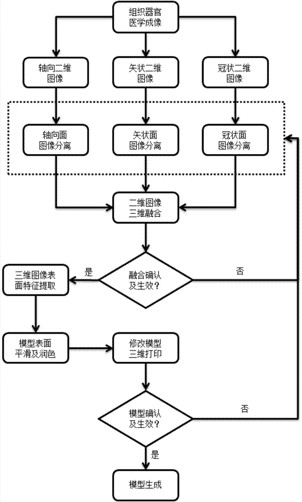
[0025] Such as figure 1 Shown, a kind of three-dimensional modeling method of the human body organ that can carry out 3D printing of the present invention comprises the following steps:

[0026] 1) Obtain medical images of tissues and organs,

[0027] The medical images of tissues and organs are obtained and stored as image files in DICOM format by means of nuclear magnetic resonance or ultrasonic scanning; wherein,

[0028] NMR:

[0029] Taking the Siemens 3T scanning device as an example, the coronal, sagittal, and axial scans are performed on the whole body of the human body or a single organ. The accuracy of each layer is 1.1×1.1mm, and the thickness of the layer is 3mm. Scanned images of human organs, which are stored in DICOM format. This method has no...


PUM


Abstract

Disclosed is a human organ three dimensional (3D) modeling method capable of performing 3D printing. The human organ 3D modeling method capable of performing the 3D printing includes: obtaining a tissue and organ medical image; simultaneously generating axial two dimensional (2D) images, sagittal 2D images and coronal 2D images of three same organs in the axial direction, the sagittal direction and the coronal direction of the tissue and organ medical image; respectively and correspondingly performing axial surface image separation, sagittal axial surface image separation and coronal axial surface image separation on the above images; performing 3D fusion of the 2D images against an image separation result; judging whether the 3D fusion takes effect or not; extracting surface features of a 3D image in medical image processing software, adding an individual file which is generated into a model, and saving the individual file as an STL formatted file; performing smoothing and polishing processing on the surface of the model; generating a file which can be printed in 3D mode and performing the 3D printing; judging whether a model which is printed out is real or not. The model which is printed out by using the human organ 3D modeling method capable of performing the 3D printing is not only realistic in surface feature, but also has an internal structure completely consistent with the structure of a human body.

Description

technical field [0001] The invention relates to a three-dimensional modeling method. In particular, it relates to a method for three-dimensional modeling of human organs capable of 3D printing. Background technique [0002] Domestically, there is no such technology for 3D modeling of formed human organs and printing them into physical models. Basically stop at 3D reconstruction, and the reconstruction technology is limited to contour reconstruction, its internal structure can not be detected, such as the patent application number 200410089299.9 3D modeling method of human limbs based on image contours, which uses a rotating conic surface to establish a 3D human limb In the model, the skeleton layer is composed of joint points and line segments connecting points to represent the skeleton structure of human limbs, and the skin layer is represented by a conic surface of rotation. Each limb only needs to adjust two deformation parameters to reflect the skin deformation of human...

Claims


Application Information

Patent Timeline
Patent Type & Authority Applications(China)
IPC IPC(8): G06T17/00
Inventor 魏程
Owner 魏程
Learn More