Uses of CHIP protein in pancreas cancer early stage diagnosis and prognosis determination
A technique for pancreatic cancer, use, and application in the field of serum markers
- Summary
- Abstract
- Description
- Claims
- Application Information
AI Technical Summary
Problems solved by technology
Method used
Image
Examples
Embodiment 1
[0029] Embodiment 1, the collection of experimental material
[0030] 1. Histopathological specimens:
[0031] The specimens of pancreatic ductal adenocarcinoma confirmed by pathological examination were collected in Peking Union Medical College Hospital from January 2006 to July 2010, and the specimens were preserved in the Peking Union Medical College Hospital Basic Surgery -80°C Specimen Bank. The inclusion and exclusion criteria of the research sample are as follows:
[0032] 1) Inclusion criteria: pathological diagnosis of pancreatic ductal adenocarcinoma specimens, cases of surgical resection.
[0033] 2) Exclusion criteria: no tumor resection, or only pancreas tissue biopsy; incomplete case data, unable to obtain follow-up information; specimens could not be prepared into immunohistochemical sections.
[0034]A total of 58 patients with pancreatic cancer were included in this study, including 25 males and 33 females, with a male-to-female ratio of 1:1.32; 38 cases wer...
Embodiment 2
[0045] Embodiment 2, experimental method
[0046] Techniques well known in the art were used to extract the histoprotein, histoprotein SDS-PAGE and Western blot, immunohistochemical detection, and ELISA to detect the expression level of CHIP in human serum. The ELISA kit of CHIP protein was purchased from Wuhan Huamei Biological Co., Ltd., kit number CBS-EL022887HU.
[0047] The judgment of immunohistochemical results is as follows:
[0048] Divided into four grades according to the percentage of stained positive cells
[0049] -Negative
[0050] +<30% positive cell count
[0051] ++30-60% positive cell count
[0052] +++>60% positive cell count
[0053] The percentage of positive cells was 60%, divided into two groups with low expression of CHIP and high expression of CHIP. Scoring was done independently by two pathologists.
[0054] Statistical Analysis:
[0055] For enumeration data, Chi-square test or Fisher's exact test was used; for univariate analysis, Log-rank ...
Embodiment 3
[0056] Embodiment 3, experimental result
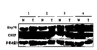
[0057] 1), SDS-PAGE and Western blot to detect the expression levels of CHIP and Hsp70 in pancreatic cancer and adjacent tissues
[0058] It was found that the expression level of CHIP in the pancreatic cancer tissues of the first and third patients was increased, and the expression level of CHIP was significantly decreased in the second and fourth patients. The expression level of Hsp70 was not significantly different, the expression level of the first and third patients increased, the expression level of the second patient had no significant difference, and the expression level of the fourth patient decreased. ( figure 1 )
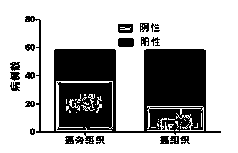
[0059] 2) Immunohistochemical detection of the expression level of CHIP protein in pancreatic cancer tissues and adjacent tissues
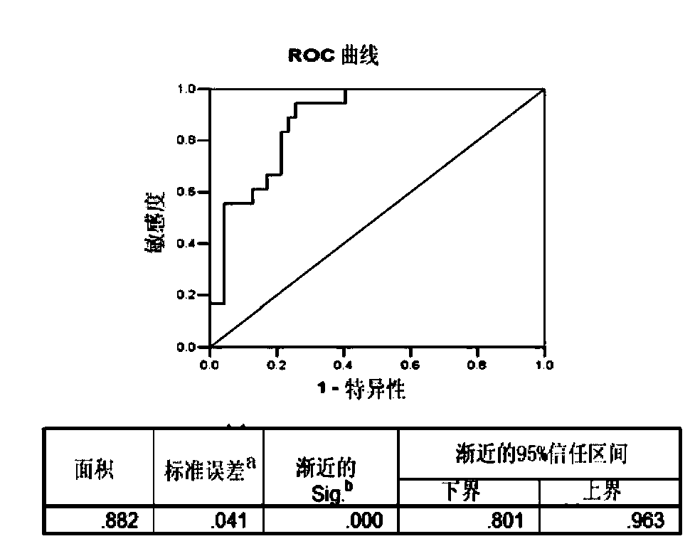
[0060] The expression of CHIP protein is mainly in the cytoplasm, which is light yellow to dark brown. There were 37 cases with negative expression of CHIP protein in paracancer...
PUM
 Abstract
Description
Claims
Application Information
 - R&D
- Intellectual Property
- Life Sciences
- Materials
- Tech Scout
- Unparalleled Data Quality
- Higher Quality Content
- 60% Fewer Hallucinations
Browse by: Latest US Patents, China's latest patents, Technical Efficacy Thesaurus, Application Domain, Technology Topic, Popular Technical Reports.
© 2025 PatSnap. All rights reserved.Legal|Privacy policy|Modern Slavery Act Transparency Statement|Sitemap|About US| Contact US: help@patsnap.com


