Methods and systems for automatically determining magnetic field inversion time of a tissue species

A technology of reversing time and organization, applied in the direction of using nuclear magnetic resonance imaging system for measurement, using magnetic variable measurement, application, etc., can solve the problem of difficult automation of the system

Inactive Publication Date: 2014-11-19
SIEMENS AG +1
View PDF4 Cites 13 Cited by
  • Summary
  • Abstract
  • Description
  • Claims
  • Application Information

AI Technical Summary

Problems solved by technology

In conventional systems, segmented cine inversion recovery sequences are used to estimate T1 and optimal TI, but these systems are difficult to automate because they are based on a series of images that differ in both contrast and cardiac phase

Method used


Image

  • Methods and systems for automatically determining magnetic field inversion time of a tissue species
  • Methods and systems for automatically determining magnetic field inversion time of a tissue species
  • Methods and systems for automatically determining magnetic field inversion time of a tissue species

Examples


Embodiment Construction

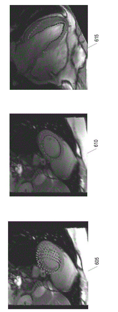
[0026] The following disclosure describes the invention in terms of several embodiments of methods, systems and apparatus intended for automatically determining tissue class zeroing reversal times. In some embodiments, the system automatically determines the zeroing reversal time using the T1 parameter map, a priori anatomical information, and T1-based tissue classification. The system can be used, for example, to improve image contrast in inversion recovery MR imaging and for workflow in late enhancement myocardial viability imaging. Additionally, the system can be used to improve scan-to-scan reliability in MRI myocardial viability imaging by automatically identifying the optimal inversion time to null the MR signal in healthy myocardium.

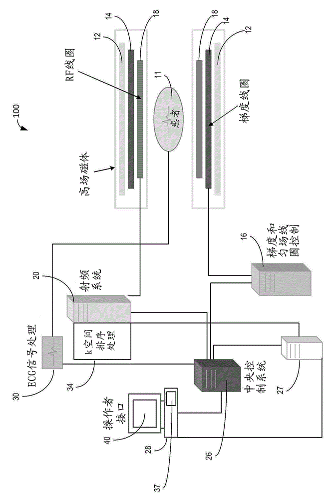
[0027] figure 1A system 100 is shown for ordering acquisitions of frequency domain components representing MR image data for storage in a k-space storage array. In system 100, magnet 12 creates a static base magnetic field within the bo...


PUM


Abstract

A computer-implemented method for determining magnetic field inversion time of a tissue species includes generating a T1-mapping image of a tissue of interest, the T1-mapping image comprising a plurality of T1 values within an expected range of T1 values for the tissue of interest. An image mask is created based on predetermined identification information about the tissue of interest. Next, an updated image mask is created based on a largest connected region in the image mask. The updated image mask is applied to the T1-mapping image to yield a masked image. Then, a mean relaxation time value is determined for the largest connected region. The mean relaxation time value is then used to determine a time point for nulling longitudinal magnetization.

Description

[0001] Cross References to Related Applications [0002] This application claims priority to US Provisional Application Serial No. 61 / 812,743, filed April 17, 2013, which is incorporated herein by reference in its entirety. technical field [0003] The present invention generally relates to methods, systems and apparatus for automatically determining magnetic reversal times for tissue classes using Tl map images, a priori information about regions of interest, and tissue classification algorithms. The disclosed methods, systems and devices may be applied, for example, to identify healthy tissue regions in cardiac applications. Background technique [0004] Conventional inversion recovery (IR) methods in Magnetic Resonance Imaging (MRI) can be used to suppress the signal from a specific tissue and thereby exploit the image contrast in adjacent tissue. For example, late enhanced IR imaging can be used to diagnose a wide range of ischemic and nonischemic cardiomyopathy. In th...

Claims


Application Information

Patent Timeline
Patent Type & Authority Applications(China)
IPC IPC(8): G01R33/50A61B5/055
CPCG06T2207/10088G06T7/0012G06T2207/30048
Inventor B.S.斯波蒂斯伍德吕晓光毕晓明薛晖C.格利尔米P.克尔曼A.格雷泽
Owner SIEMENS AG
Why Eureka
Try Eureka