High content image flow biological microscopic analysis system

A technology of microscopic analysis and image flow, applied in the field of flow cytometry, can solve problems such as difficult to provide quantitative and statistical data of cell populations, limited number of cells, etc.

Inactive Publication Date: 2015-01-07
SHANGHAI JIAO TONG UNIV
View PDF6 Cites 21 Cited by
  • Summary
  • Abstract
  • Description
  • Claims
  • Application Information

AI Technical Summary

Problems solved by technology

To obtain images of cells, researchers must use a microscope to observe, but the number of cells that can be observed with a microscope is very limited, and it is difficult to provide quantitative and statistical data on cell populations
Therefore, using traditional cell analysis techniques, we are faced with the dilemma that there is no technique that can provide both statistical data of cell populations and obtain cell images

Method used


Image

  • High content image flow biological microscopic analysis system
  • High content image flow biological microscopic analysis system

Examples


Embodiment Construction

[0034] The present invention will be described in detail below in conjunction with specific embodiments. The following examples will help those skilled in the art to further understand the present invention, but do not limit the present invention in any form. It should be noted that those skilled in the art can make several modifications and improvements without departing from the concept of the present invention. These all belong to the protection scope of the present invention.

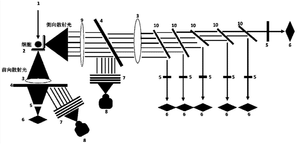
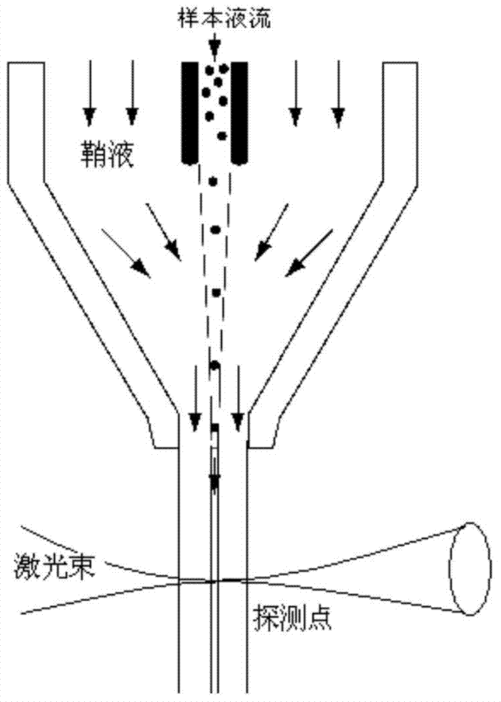
[0035] The high-content image flow biomicroscopic analysis system provided by the present invention is mainly composed of several parts such as a liquid flow system, an optical system, an electronic system, and a flow chamber.

[0036] The liquid flow system includes a flow chamber. The liquid flow system injects the sample cell suspension and the system sheath liquid into the flow chamber. The flow chamber is used to make the cells focus on the center of the liquid flow under the constraint of the...


PUM


Abstract

The invention provides a high content image flow biological microscopic analysis system. The system comprises a liquid flow system and an optical system, wherein the liquid flow system enables cells in sample cell suspension to be focused in the center of a liquid flow under the constraint of the system sheath liquid flow and to flow through a detection window of a flow chamber one by one; the optical system comprises optical sources, an optical path system and an optical filter stack; the optical sources include a halogen lamp and a laser; through the optical path system and the optical filter stack formed by a plurality of dichroscopes, the optical system generates a bright field cell image, a six-channel fluorescent cell image and six PMT (photomultiplier tube) optical intensity signals with different wave bands. The high content image flow biological microscopic analysis system integrates flow cytometry and fluorescence microscopic imaging, has a plurality of detection channels, can collect the cell images of the cells passing through the flow chamber one by one and can carry out quantitative analysis on each cell image by utilizing analysis software, thus providing the statistical data of a cell group and the information of the cell morphology, cell structures and subcellular signal distribution.

Description

technical field [0001] The invention relates to flow cytometry, in particular to a high-content image flow biomicroscopic analysis system. Background technique [0002] Flow cytometry is a technique for rapid quantitative analysis and sorting of cells or other biological particles (such as microspheres, bacteria, small model organisms, etc.) arranged in a single row in a liquid flow. Using traditional flow cytometry technology, researchers can analyze tens of thousands of cells and obtain the numerical value of scattered light signal and fluorescent signal of each cell, so as to obtain various statistical data of cell population and find rare cells subgroup. [0003] However, traditional flow cytometry technology still has limitations, that is, the obtained cell information is very limited. For researchers, a cell is just a point on a scatter plot, not a real cell image, lacking information about cell morphology, cell structure, and signal distribution at the subcellular l...

Claims


Application Information

Patent Timeline
Patent Type & Authority Applications(China)
IPC IPC(8): G01N15/14
Inventor 胡洁戚进刘超
Owner SHANGHAI JIAO TONG UNIV
Why Eureka
Try Eureka