Cell detection method based on sliding window and depth structure extraction features

A sliding window and feature extraction technology, applied in character and pattern recognition, instruments, computer parts, etc., can solve the problem of inability to deal with multiple changes in cell shape and different scales

Active Publication Date: 2015-02-11
NANJING UNIV OF INFORMATION SCI & TECH
View PDF0 Cites 29 Cited by
  • Summary
  • Abstract
  • Description
  • Claims
  • Application Information

AI Technical Summary

Problems solved by technology

[0006] The technical problem to be solved by the present invention is to overcome the problem that the existing segmentation-based detection algorithm cannot handle pathological slice cells due to multiple changes in cell shape and different scales, and to provi...

Method used


Image

  • Cell detection method based on sliding window and depth structure extraction features
  • Cell detection method based on sliding window and depth structure extraction features
  • Cell detection method based on sliding window and depth structure extraction features

Examples


Embodiment Construction

[0033] Below in conjunction with accompanying drawing, technical scheme of the present invention is described in further detail:

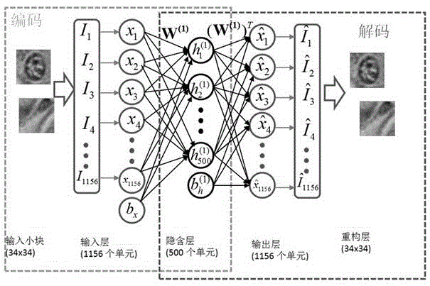
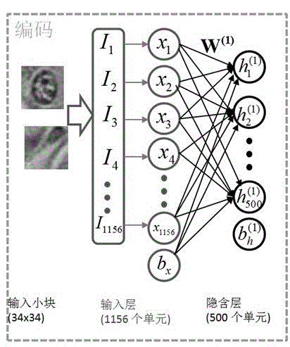
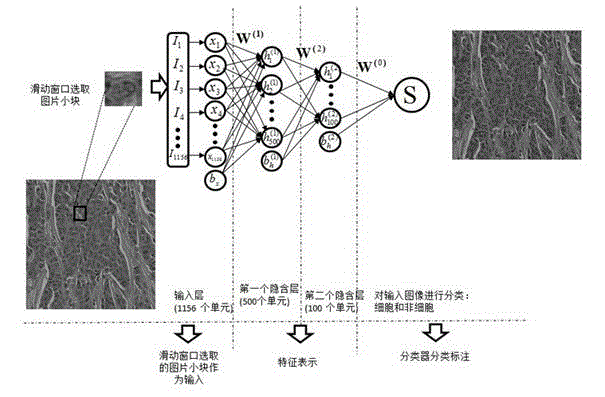
[0034] A cell detection method based on sliding window and deep structure extraction features of the present invention, such as image 3 shown, including the following steps:

[0035] Step 1. Selection of training samples: select small blocks containing cells and non-cellular small blocks in the pathological image, wherein the non-cellular small blocks include small blocks with some cells and small blocks that do not contain cells at all;

[0036] Regarding the selection of cell blocks, clinicians with professional pathological knowledge will mark them in the large-scale slice images, and the program will intercept these marked points to the original image to capture a small square block with the marked point as the center and a side length of 34 pixels. . These cell-containing clumps serve as cell positive samples. For large cells, take a 50 p...


PUM


Abstract

The invention discloses a cell detection method based on a sliding window and depth structure extraction features. The cell detection method is used for automatically detecting cells by utilizing depth model extraction features and then applying a sliding window technology to a pathological section image. The cell detection method comprises the following steps: section image blocking, training of stacked and sparse self-coding of a feature extraction model, detector training, scanning of a large image by the sliding window and cell position labeling. According to the cell detection method, the large section image is used as a search object, the positions of cells in the image can be found more accurately, faster and completely by adopting a new method of combining a detector and the sliding window, and a good detection effect can be achieved for some unobvious cells in the image. The automatic cell detection method disclosed by the invention can be used for assisting a clinical doctor in carrying out quantitative evaluation on digital pathological sections and accurately and rapidly carrying out clinical diagnosis, so that the diagnosis difference of different observers or one observer at different time periods is reduced.

Description

technical field [0001] The invention discloses a cell detection method based on a sliding window and a depth structure extraction feature, and relates to the technical field of image information processing. Background technique [0002] With the generation of digital scanning technology for large slice images and the improvement of scanning efficiency, the digital display and storage of histopathological slides has become realistic and feasible. Higher quality analysis of pathological images is possible with digital technology. Because the characteristics of various cancer cells and tissues can be found from the pathological imaging images of tissue slices, and can be used to assist doctors in diagnosis, but there are still few technical researches on medical image processing. Analysis tools for pathological images are very important. [0003] A large part of the research on histopathological images focuses on the resolution of specific tissue structures, such as lymphocyt...

Claims


Application Information

Patent Timeline
IPC IPC(8): G06K9/46G06K9/62
CPCG06V20/695G06V10/44G06F18/24147
Inventor 徐军项磊
Owner NANJING UNIV OF INFORMATION SCI & TECH
Why Eureka
Try Eureka