Blind source separation method for extracting J wave signals in electrocardiogram

A blind source separation and electrocardiogram technology, which is used in medical science, sensors, diagnostic recording/measurement, etc., to reduce the amount of calculation, improve the separation accuracy, and improve the separation accuracy.

Active Publication Date: 2015-02-25
TAIYUAN UNIV OF TECH
View PDF3 Cites 11 Cited by
  • Summary
  • Abstract
  • Description
  • Claims
  • Application Information

AI Technical Summary

Problems solved by technology

[0003] The present invention provides a blind source separation method for extracting J wave signals in el

Method used


Image

  • Blind source separation method for extracting J wave signals in electrocardiogram
  • Blind source separation method for extracting J wave signals in electrocardiogram
  • Blind source separation method for extracting J wave signals in electrocardiogram

Examples


Embodiment Construction

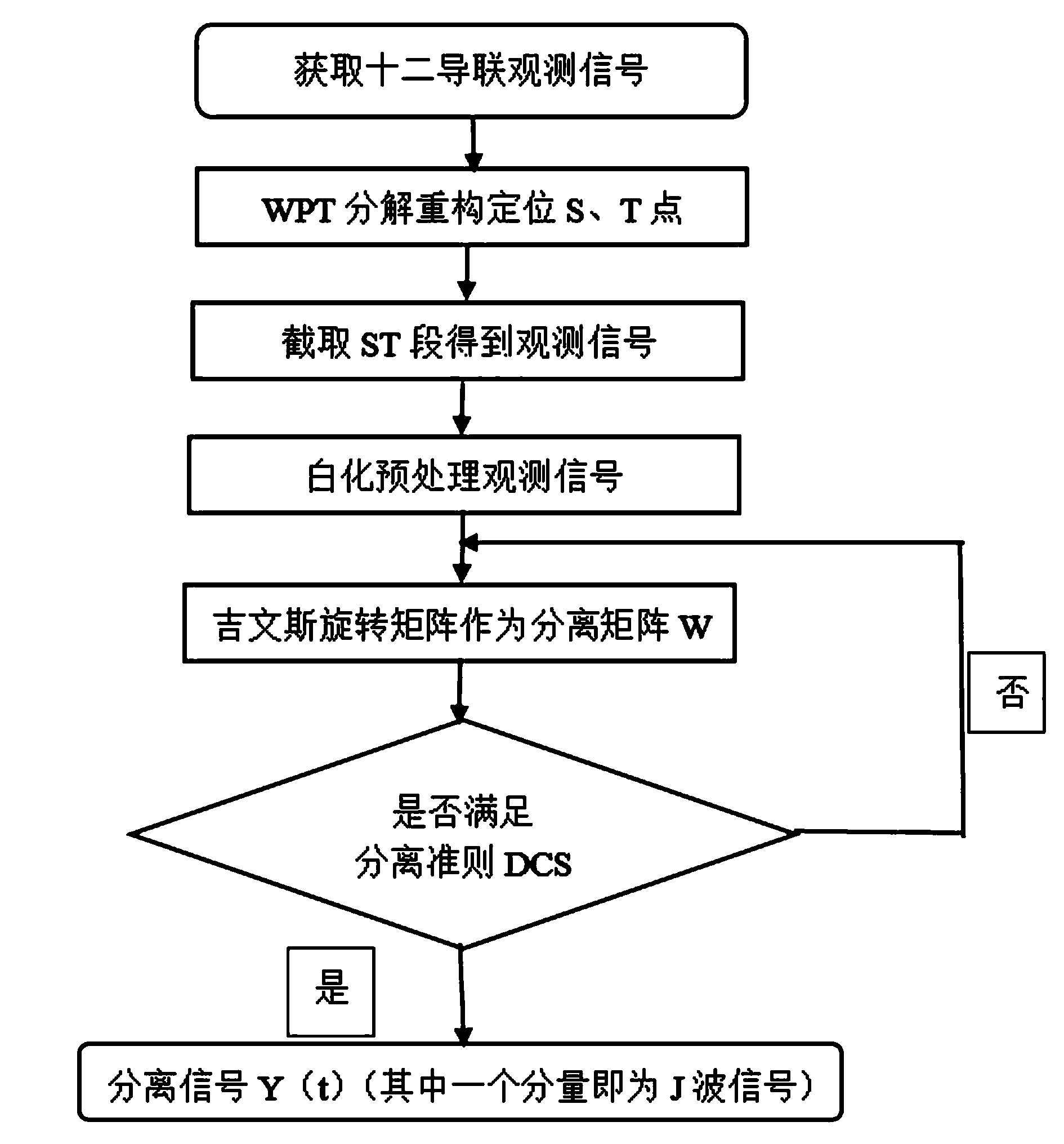
[0017] A blind source separation method for extracting J-wave signals in electrocardiograms, comprising the following steps:

[0018] 1) Obtain the twelve-lead ECG of patients with J wave syndrome as the initial observation signal;

[0019] 2) Extract the new observation signal containing J wave from the initial observation signal: select db3 as the wavelet function ψ(t), and its corresponding scaling function is φ(t), and the wavelet function ψ(t) and scaling function φ(t ) to calculate the filter H 0 (z) and H 1 (z) filter coefficient h 0 (k) and h 1 (k),

[0020] h 0 ( k ) = φ 1,0 ( t ) ...


PUM


Abstract

The invention relate to a J wave extraction method, in particular to a blind source separation for extracting J wave signals in an electrocardiogram on the basis of a cyclostationarity standard. According to the method, a Givens rotation matrix serves as a separation matrix. The J wave signals are a new index of upper chamber repolarization of the electrocardiogram (ECG), and it is indicated in clinical data that shapes of the J wave signals in the ECG are different, randomness is high and the J wave signals are lost in the normal ECG and can not be observed sometimes. In order to accurately extract the J wave signals, according to the method, wavelet packet transform (WPT) is adopted in advance to analyze, reconstruct and position an ST section where J waves are located, and accordingly the calculation amount of blind source separation (BSS) is reduced; according to the characteristics of the ECG and the J wave signals, the ECG stable in circulation and the non-cycle J wave signals are effectively separated by means of the BSS method based on higher-order cyclic cumulant, and guarantees are provided for research of the J wave signals in a time domain in the next step and separation of the good state and the high-risk state.

Description

technical field [0001] The invention relates to a J-wave extraction method, in particular to a blind source separation method for extracting J-wave signals in an electrocardiogram based on a cyclostationarity criterion with a Givens rotation matrix as a separation matrix. Background technique [0002] The J wave belongs to a normal ECG (cardiac signal) variation, but when the shape, duration and amplitude of the J wave change significantly, such as: the J point following the QRS wave shifts from the baseline for a duration > 0.03s, Amplitude > 0.05mv, forming peak-shaped, hump-shaped, dome-shaped waveforms or ST-segment elevation may indicate malignant arrhythmia events such as ventricular tachycardia and ventricular fibrillation, and even lead to sudden cardiac death. In recent years, as a new indicator of ECG ventricular repolarization, the mutated J wave has been paid attention to by the medical community. Therefore, it is of great clinical significance to distingui...

Claims


Application Information

Patent Timeline
IPC IPC(8): A61B5/0402
CPCA61B5/318
Inventor 赵菊敏朱贝贝李灯熬
Owner TAIYUAN UNIV OF TECH
Why Eureka
Try Eureka