
Method and device for amplifying ultrasound image

A technology of ultrasound image and magnification device, which is applied in the field of image processing, can solve problems such as blurring of ultrasound images and generation of artifacts, and achieve the effect of meeting medical clinical requirements

Active Publication Date: 2015-03-25
EDAN INSTR
View PDF4 Cites 11 Cited by
  • Summary
  • Abstract
  • Description
  • Claims
  • Application Information

AI Technical Summary

Problems solved by technology

[0007] In view of this, an embodiment of the present invention provides a method and device for magnifying an ultrasonic image to solve the problem that the existing ultrasonic image magnifying method causes the enlarged ultrasonic image to be blurred and generate artifacts, so that the enlarged ultrasonic image can maintain the original image the details

Method used


Image

  • Method and device for amplifying ultrasound image
  • Method and device for amplifying ultrasound image
  • Method and device for amplifying ultrasound image

Examples


Embodiment 1

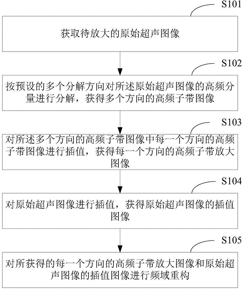
[0069] figure 1 The first implementation flow of the ultrasonic image magnification method provided by Embodiment 1 of the present invention is shown. For the convenience of description, only the parts related to the present invention are shown.

[0070] Such as figure 1 As shown, the method includes:

[0071] In step S101, the original ultrasound image to be enlarged is acquired.

[0072] In the embodiment of the present invention, the original ultrasonic image after logarithmic compression, dynamic range transformation and digital scan transformation is pre-acquired. After the original ultrasound image is collected, the user's selection instruction is received, and the image area corresponding to the selection instruction (ie ROI image, Region of Interesting) is obtained, which is defined as the original ultrasound image to be enlarged; or the ROI is automatically selected according to the preset selection instruction image. Since the enlarged image will replace the orig...

Embodiment 2

[0132] Figure 10 The structure of the ultrasonic image magnification device provided by Embodiment 2 of the present invention is shown, and for the convenience of description, only the parts related to the present invention are shown.

[0133] Such as Figure 10 As shown, the device includes:

[0134] The acquiring module 11 is configured to acquire the original ultrasonic image to be enlarged.

[0135] The decomposition module 12 is configured to decompose the high-frequency components of the original ultrasonic image in multiple directions according to a plurality of preset decomposition images, and obtain high-frequency sub-band images in multiple directions.

[0136] An interpolation module 13, configured to interpolate the high-frequency sub-band images in each direction among the high-frequency sub-band images in each direction, obtain the enlarged high-frequency sub-band images in each direction, and perform interpolation on the original ultrasonic image , to obtain...


PUM


Abstract

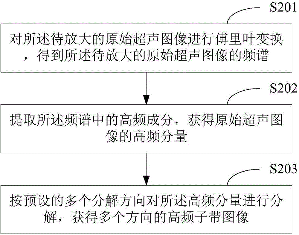
The invention belongs to the technical field of image processing, and provides a method and device for amplifying an ultrasound image. The method includes the steps that an original ultrasound image to be amplified is acquired; the high-frequency component of the original ultrasound image is decomposed according to a plurality of preset decomposition directions, so that high-frequency sub-band images in multiple directions are acquired; interpolation is conducted on the high-frequency sub-band image in each direction in the high-frequency sub-band images in the multiple directions, and the high-frequency sub-band amplified image in each direction is acquired; interpolation is conducted on the original ultrasound image, and an interpolation image of the original ultrasound image is acquired; frequency domain reconstruction is conducted on the acquired high-frequency sub-band amplified image in each direction and the acquired interpolation image of the original ultrasound image, and accordingly the amplified ultrasound image is acquired. The amplified ultrasound image can keep the details of the original image, ghosting-artifacts can be avoided, the medical clinic requirement can be met, and doctor diagnosis can be facilitated.

Description

technical field [0001] The invention belongs to the technical field of image processing, and in particular relates to an ultrasonic image enlargement method and device. Background technique [0002] The purpose of image upscaling is to obtain high-resolution images from low-resolution images, that is, super-resolution reconstruction. Super-resolution ultrasound image reconstruction technology has important application value in clinical medicine. There are three types of existing super-resolution image generation methods, namely: [0003] 1. Interpolation of single-frame low-resolution images: such as classic linear interpolation and cubic interpolation. This kind of amplification method is simple and easy to implement and widely used, but it is easy to cause the interpolated image to become blurred. [0004] 2. Reconstruction of multi-frame low-resolution images: such as iterative back projection (IBP, Iterative Back Projection), convex set projection (POCS, Projections On...

Claims


Application Information

Patent Timeline
Patent Type & Authority Applications(China)
IPC IPC(8): G06T3/40
CPCG06T3/4007G06T3/4084
Inventor 王明悦
Owner EDAN INSTR
Learn More