Automatic tumor image region segmentation method based on improved level set

A technology of tumor area and tumor imaging, which is applied in the field of image processing, can solve the problem that it is difficult to automatically distinguish between tumor area and bladder area, and achieve the effect of strong robustness, fast speed and high precision

Active Publication Date: 2018-04-13
INST OF AUTOMATION CHINESE ACAD OF SCI
View PDF2 Cites 0 Cited by
  • Summary
  • Abstract
  • Description
  • Claims
  • Application Information

AI Technical Summary

Problems solved by technology

[0004] The invention provides an automatic tumor image region segmentation method based on an improved level set, which is used to solve the problem that it is difficult to automatically distinguish the tumor region and the bladder region in cervical cancer tumor segmentation, so that users can quickly and accurately segment cervical cancer tumors to assist Surgeon conducts diagnosis, treatment and efficacy evaluation

Method used


Image

  • Automatic tumor image region segmentation method based on improved level set
  • Automatic tumor image region segmentation method based on improved level set
  • Automatic tumor image region segmentation method based on improved level set

Examples


Embodiment Construction

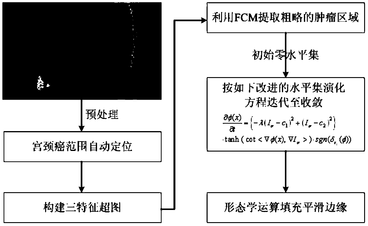
[0017] figure 1 It is a flow chart of the automatic tumor image region segmentation method based on the improved level set of the present invention, such as figure 1 As shown, the automatic tumor image region segmentation method based on the improved level set of the present invention includes:

[0018] S1. Acquiring the original PET image containing the lesion area to be segmented and performing preprocessing and positioning to determine the preprocessed PET image of the lesion area to be segmented;

[0019] Preferably, the acquisition of the original PET image containing the lesion area to be segmented and performing preprocessing and positioning so as to determine the preprocessed PET image of the lesion area to be segmented comprises:

[0020] Divide the voxel gray value in the original PET image containing the lesion area by the injected contrast agent dose and the patient's body weight to convert it into an SUV value, and then perform Gaussian filtering and up-sampling,...


PUM


Abstract

The present invention provides an automatic tumor image region segmentation method based on an improved level set, comprising: acquiring an original PET image including a lesion area to be segmented and performing preprocessing and positioning to determine the preprocessed PET image of a lesion area to be segmented; Construct a hypergraph according to the CT image of the lesion area and the PET image of the lesion area to be segmented after the preprocessing, thereby preliminarily determining the rough tumor area in the PET image as an initial zero level set; performing an improved level on the initial zero level set A set method is used to determine the tumor area; edge smoothing is performed on the tumor area according to the morphological operation. The method of the invention can realize rapid and accurate segmentation of tumor regions, thereby assisting surgeons in diagnosis, treatment and curative effect evaluation.

Description

technical field [0001] The invention relates to image processing technology, in particular to an automatic tumor image region segmentation method based on an improved level set. Background technique [0002] Cervical cancer is one of the three most common malignant tumors of female reproductive organs, and one of the main malignant tumors that threaten women's life and affect the quality of life, ranking first among female reproductive organ malignant tumors. According to the 2014 World Cancer Report published by The International Agency for Research on Cancer under the World Health Organization at its headquarters in Lyon, France on February 3, the number of new cases of cervical cancer worldwide in 2012 reached more than 500,000. Among female malignant tumors, it ranks fourth after breast cancer, rectal cancer, and lung cancer. Over the same period, cervical cancer caused more than 260,000 deaths, and its mortality rate is second only to breast cancer, lung cancer, and rec...

Claims


Application Information

Patent Timeline
Patent Type & Authority Patents(China)
IPC IPC(8): G06T7/136
Inventor 田捷牟玮陈喆杨凤
Owner INST OF AUTOMATION CHINESE ACAD OF SCI
Why Eureka
Try Eureka