Optical microangiography image segmentation and evaluation method

A technology for contrast images and microvessels, which is applied in image analysis, image enhancement, image data processing, etc., and can solve problems such as inapplicable images

Active Publication Date: 2015-09-02
ZHEJIANG UNIV
View PDF4 Cites 6 Cited by
  • Summary
  • Abstract
  • Description
  • Claims
  • Application Information

AI Technical Summary

Problems solved by technology

However, the selection of the global threshold has been a more difficult problem
In the current image segmentation algorithm, some existing threshold segmentati

Method used


Image

  • Optical microangiography image segmentation and evaluation method
  • Optical microangiography image segmentation and evaluation method
  • Optical microangiography image segmentation and evaluation method

Examples


Embodiment Construction

[0050] The present invention will be further described below in conjunction with the accompanying drawings and implementation examples.

[0051] A method for segmenting an optical microangiography image, the method specifically comprising the following steps:

[0052] Establish the mathematical statistics of optical microangiography based on the "stochastic vector sum of phase amplitude" model:

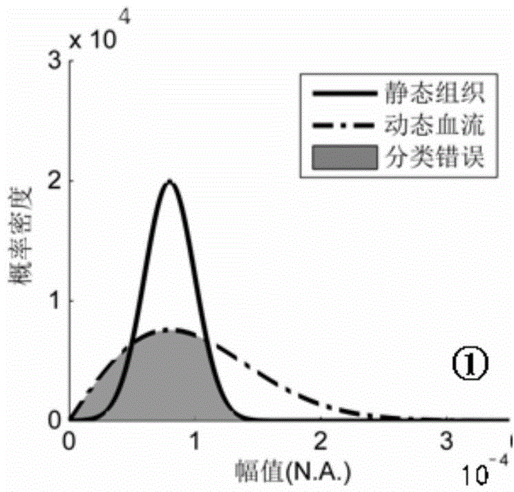
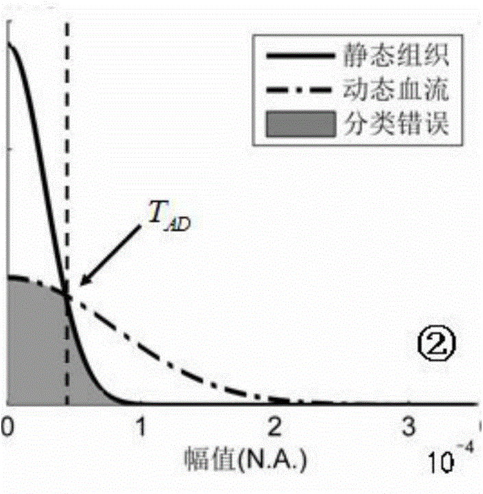
[0053] Applying the "random phase amplitude vector sum" model in statistical optics, the OCT (Optical Coherence Tomography) complex-valued signal A(z,x,t) at a certain point in the sample space domain is represented as multiple independent tiny The sum of the contributions of the backscattered light from the scattered particles, that is, the complex superposition of multiple small independent phase amplitude vectors;

[0054] For the dynamic blood flow area, the moving red blood cells are independent tiny scatterers, and the optical scattering signals of the independent tiny scattere...


PUM


Abstract

The invention discloses an optical microangiography image segmentation and evaluation method. Based on optical coherence tomography (OCT), unmarked and three-dimensional optical microangiography needs to split a dynamic flood flow signal from a static organization background so as to realize three-dimensional radiography of a capillary. In the invention, ''a random phase vector'', a statistical optical model and a difference algorithm of optical microangiography (comprising an amplitude difference and a complex signal difference) are combined so that a signal statistics theory model of the optical microangiography is firstly derived and a statistics characteristic difference of the dynamic flood flow signal and the static organization background is successfully interpreted, wherein the statistics characteristic difference of the dynamic flood flow signal and the static organization background is a contrast source of the microangiography. Therefore, an automatic threshold segmentation algorithm based on a minimum segmentation error rate and an objective evaluation standard based on a residual segmentation error rate are proposed.

Description

technical field [0001] The present invention relates to optical coherence tomography (Optical Coherence Tomography, OCT) and optical microangiography (OCT Angiography, Angio-OCT) technology, especially to a kind of optical microangiography image based on "random phase amplitude vector sum" statistical model Segmentation and evaluation methods. technical background [0002] Compared with the current biomedical imaging methods, OCT imaging technology has the advantages of label-free, non-contact, non-invasive, real-time, high sensitivity and high resolution. Rapid development, and has been widely accepted clinically. The OCT system mainly obtains the refractive index information of the sample by detecting the change of the backscattered light intensity caused by the optical inhomogeneity of the biological sample, and then obtains the optical structure image of the sample. However, in the early stage of the disease, the difference in scattering characteristics between normal ...

Claims


Application Information

Patent Timeline
IPC IPC(8): G06T7/00
CPCG06T7/0012G06T7/10G06T2207/30101
Inventor 李鹏丁志华程宇轩郭立潘聪
Owner ZHEJIANG UNIV
Why Eureka
Try Eureka