Eliminating motion effects in medical images caused by physiological function

A technology in medical images and images, applied in image data processing, ultrasonic/sonic/infrasonic image/data processing, image enhancement, etc.

Active Publication Date: 2015-10-07
KONINKLJIJKE PHILIPS NV
View PDF17 Cites 6 Cited by
  • Summary
  • Abstract
  • Description
  • Claims
  • Application Information

AI Technical Summary

Problems solved by technology

But identifying and removing images from the sequence is a manual process that requires individual examination and evaluation of each image and its view of the diaphragm

Method used


Image

  • Eliminating motion effects in medical images caused by physiological function
  • Eliminating motion effects in medical images caused by physiological function
  • Eliminating motion effects in medical images caused by physiological function

Examples


Embodiment Construction

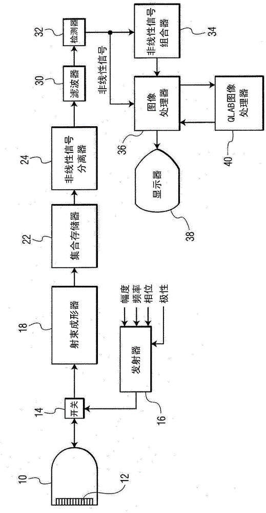
[0019] first reference figure 1 , shows in block diagram form an ultrasound system constructed in accordance with the principles of the present invention. The system operates by scanning a two-dimensional or three-dimensional region of the body being imaged using ultrasound emission beams. As each beam is emitted through the body along its guided path, the beam returns an echo signal having linear and non-linear (fundamental and harmonic frequencies) components corresponding to the frequency components emitted. The transmit signal is modulated by the non-linear response of contrast agent microbubbles encountered by the beam, generating echo signals with harmonic components.

[0020] figure 1 The ultrasound system in ® utilizes a transmitter 16 that emits waves or pulses of selected modulation characteristics in a desired beam direction to return harmonic echo components from scatterers within the body. The transmitter responds to a number of control parameters that determin...


PUM


Abstract

Ultrasonic imaging methods are described for eliminating the effects of physiological motion such as respiratory motion from image data of a sequence of images which is to be used for quantified analysis such as time-intensity curve computation. A bright reflector in the images of the loop which exhibits motional effects such as cyclical variation in brightness or position from image to image is identified and its variation used to identify cycles of motion. A phase or range of phases of the cycles is identified. Images acquired during the identified phase or phase range of the motion cycles during the loop are then used for quantified processing.

Description

[0001] Cross References to Related Applications [0002] This application claims priority to US Provisional Application Serial No. 61 / 753898, filed January 17, 2013, which is hereby incorporated by reference. technical field [0003] The present application relates to an ultrasonic diagnostic imaging system, and in particular, to the use of an ultrasonic diagnostic imaging system to evaluate the progress of therapeutic treatment of a tumor. Background technique [0004] International Patent Publication WO 2006 / 090309 (Bruce et al.) describes an ultrasound imaging technique for detecting lesions in the liver by using ultrasound contrast agents. A bolus of contrast medium is introduced into the body and the time it takes for the contrast medium to reach the liver is measured. When a bolus of contrast agent travels through the blood vessels of the body and begins to appear at a specific organ or location in the body, the establishment of contrast in the image is called "washin...

Claims


Application Information

Patent Timeline
Patent Type & Authority Applications(China)
IPC IPC(8): A61B8/06A61B8/08
CPCA61B8/06A61B8/481A61B8/483A61B8/5223A61B8/5276A61B8/5284G06T7/246G16H50/30G06T7/0012G06T2207/10004G06T2207/30096
Inventor M·阿韦里基乌D·克里斯托菲迪斯
Owner KONINKLJIJKE PHILIPS NV
Why Eureka
Try Eureka