Method and system for automatically scanning intracranial cerebral vessels

An automatic scanning and cerebrovascular technology, applied in the field of cerebrovascular positioning and scanning, can solve the problems of lack of cerebral blood flow monitoring function and automatic adjustment function

Active Publication Date: 2015-11-11
SHENZHEN DELICA MEDICAL EQUIP CO LTD
View PDF7 Cites 14 Cited by
  • Summary
  • Abstract
  • Description
  • Claims
  • Application Information

AI Technical Summary

Problems solved by technology

Likewise, the product also lacks cerebral blood flow monitoring and autoregulation

Method used


Image

  • Method and system for automatically scanning intracranial cerebral vessels
  • Method and system for automatically scanning intracranial cerebral vessels
  • Method and system for automatically scanning intracranial cerebral vessels

Examples


Embodiment 1

[0063] Example 1 of the positioning process: locating the left middle cerebral artery (LMCA)

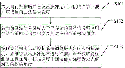
[0064] The surface of the probe is coated with couplant, placed on the left temporal window, and the initial state is perpendicular to the temporal window; the scanning depth is adjusted to 30mm; the scanning is turned on, and the probe starts to rotate; the signal strength is judged in real time during the scanning process, and when a strong signal is found, record the probe Angle; continue to scan, if there is a stronger signal, record the new probe angle; until the scan is completed, record the probe angle at the strongest signal. Increase the scanning depth by 5mm to 35mm, repeat the above process, and record the probe angle at the strongest signal. Thereafter, the scanning depth was increased by 5mm each time until it was at 55mm, and the probe angle at each scanning depth was recorded. Through the above scanning, the sequence of probe angles completely covers the LMCA, and the...

Embodiment 2

[0065] Example 2 of the positioning process: Locating the left anterior cerebral artery (LACA)

[0066] The surface of the probe is coated with couplant, placed on the left temporal window, and the initial state is perpendicular to the temporal window; the scanning depth is adjusted to 55mm; the scanning is turned on, and the probe starts to rotate; the signal strength is judged in real time during the scanning process, and when a strong signal is found, record the probe Angle, determine whether the position is tilted forward and upward; if so, continue scanning, if there is a stronger signal, record the new probe angle, and determine whether the position is tilted forward and upward; until the scan is completed, record the probe at the position with the strongest signal angle. Increase the scanning depth by 5mm to 60mm, repeat the above process, and record the position of the probe with the strongest reverse signal. Thereafter, the scanning depth was increased by 5mm each ti...

Embodiment 3

[0067] Example 3 of the positioning process: positioning the left posterior cerebral artery (LPCA)

[0068] The surface of the probe is coated with couplant, placed on the left temporal window, and the initial state is perpendicular to the temporal window; the scanning depth is adjusted to 55mm; the scanning is turned on, and the probe starts to rotate; the signal strength is judged in real time during the scanning process, and when a strong signal is found, record the probe Angle, to determine whether the position is tilted backwards and upwards; if so, continue scanning, if there is a stronger signal, record the new probe angle, and determine whether the position is tilted backwards and upwards; until the scanning is completed, record the probe at the position with the strongest signal angle. Increase the scanning depth by 5mm to 60mm, repeat the above process, and record the position of the probe with the strongest reverse signal. Thereafter, the scanning depth was increas...


PUM


Abstract

The invention discloses a method and system for automatically scanning intracranial cerebral vessels. The method includes: adjusting probe angles and scanning depths, and performing scanning to acquire the probe angles corresponding to maximum echo signal strength when a cerebral vessel to be scanned is in each scanning depth; deleting the repeated probe angles to obtain a probe angle sequence; according to the probe angle sequence, performing scanning until all the probe angles in the probe angle sequence are covered; during acquiring the probe angles and scanning according to the probe angle sequence, receiving a current echo signal corresponding to the current probe angle in real time, acquiring the corresponding current echo signal strength in real time, judging a signal display color of the current echo signal strength according to an echo signal strength interval-signal display color list, and displaying the color on a corresponding display point in real time. The probe angle sequence is acquired first, real-time scanning is then performed according to the probe angle sequence, real-time point scanning is performed on a display screen, and visually acquiring information is facilitated for users.

Description

technical field [0001] The invention relates to the technical field of cerebrovascular positioning and scanning, in particular to a method and system for automatically scanning intracranial cerebrovascular. Background technique [0002] It is a known method to fix the ultrasound probe with a certain device so that the ultrasound can scan the intracranial blood vessels. However, the current methods have certain deficiencies in terms of the convenience of operation or the complexity of the device itself. Some of the existing installations are as follows: [0003] (1) The Cerevast company in the United States has developed a device that uses an array of ultrasound probes (the left and right temporal windows each have 6 ultrasound probe array elements, and the occipital window has four ultrasound probe array elements, all of which are placed in fixed positions outside the skull) ) to ensure the coverage of the ultrasonic signal on the patient, while reducing the dependence o...

Claims


Application Information

Patent Timeline
Patent Type & Authority Applications(China)
IPC IPC(8): A61B8/06
Inventor 王筱毅王江涛
Owner SHENZHEN DELICA MEDICAL EQUIP CO LTD
Why Eureka
Try Eureka