Method and apparatus for detecting breast muscle in breast image

A technology for chest muscles in images, which is applied in the field of chest muscle detection, can solve problems such as the difficulty in detecting the area where the chest muscles are located, the inconsistent gray value of the breast image area, and the breast image does not meet the clinical needs, etc., achieving high accuracy, fast speed, The effect of improving performance

Active Publication Date: 2016-03-30
SHANGHAI UNITED IMAGING HEALTHCARE
View PDF4 Cites 10 Cited by
  • Summary
  • Abstract
  • Description
  • Claims
  • Application Information

AI Technical Summary

Problems solved by technology

[0003] In breast image processing, it is necessary to detect the area where the pectoral muscles are located. However, the current method is usually to post-process the original breast images collected by FFDM. For example, the chest muscles are detected after the thickness and tissue are balanced. This is Because the contrast between different tissues in the original breast image is relatively poor, it is difficult to detect the area where the pectoralis is located
However, performing pectoral muscle detection on the post-processed original breast image will make it impossible to use the information of the pectoral muscle area or the information of the pectoral muscle area will cause certain interference to the above-mentioned post-processing process during the post-processing process. As a result, the final obtained breast image may not meet the actual clinical needs, such as the poor contrast of the breast image, the inconsistency of the gray value between the breast image area and the breast edge, etc., which also reduces the performance of the FFDM system

Method used


Image

  • Method and apparatus for detecting breast muscle in breast image
  • Method and apparatus for detecting breast muscle in breast image
  • Method and apparatus for detecting breast muscle in breast image

Examples


Embodiment 1

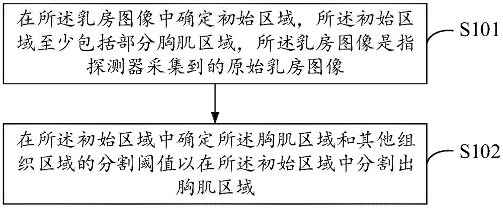
[0091] See figure 1 , figure 1 It is a flow chart of the method for detecting pectoral muscles in breast images according to Embodiment 1 of the present invention, such as figure 1 As shown, in this embodiment, the method for detecting pectoral muscles in the breast image includes:

[0092] S101: Determine an initial area in the breast image, the initial area includes at least a part of the pectoral muscle area, and the breast image refers to the original breast image collected by the detector;

[0093] S102: Determine a segmentation threshold of the pectoral muscle region and other tissue regions in the initial region to segment a pectoral muscle region in the initial region.

[0094] Execute S101, roughly segment the acquired original breast image, and determine an initial area including at least part of the pectoral muscle area. Those skilled in the art know that when the breast image (the original breast image collected by the detector, usually including horizontal boun...

Embodiment 2

[0138] Different from Embodiment 1, in this embodiment, in order to further improve the accuracy of the detected pectoral muscle region, the positional relationship between the pectoral muscle wall boundary line of the first region segmented based on the segmentation threshold and the pectoral muscle wall boundary line of the initial region Use judgment accordingly to determine the final pectoral region. See Figure 13 , Figure 13 It is a flow chart of the method for detecting pectoral muscles in breast images according to Embodiment 2 of the present invention, such as Figure 13 As shown, the method for detecting pectoralis in the breast image comprises:

[0139] S201: Determine an initial area in the breast image, the initial area includes at least a part of the pectoral muscle area, and the breast image refers to the original breast image collected by the detector;

[0140] S202: Determine a segmentation threshold of the pectoralis region and other tissue regions in the...


PUM


Abstract

The invention discloses a method and an apparatus for detecting breast muscle in a breast image. The method for detecting the breast muscle in the breast image comprises: determining an initial region in the breast image, wherein the initial region at least comprises a partial breast muscle region, and the breast image refers to an original breast image acquired by a detector; and determining a segmentation threshold of the breast muscle region and other tissue regions in the initial region to segment out a breast muscle region in the initial region. According to the technical scheme, the accuracy of detecting the breast muscle region is high and the speed of detecting the breast muscle in an actual application process is high; and in addition, the performance of an FFDM system is improved while the quality of the finally obtained breast image is improved to a certain extent.

Description

technical field [0001] The invention relates to the technical field of image processing, in particular to a method and device for detecting pectoral muscles in breast images. Background technique [0002] With the development of computer science and information technology, medical imaging technology has also been developed rapidly, and various medical imaging systems are emerging. Full-field digital mammography (FFDM, full-field digital mammography) system and digital breast tomography (DBT, digital breasttomosynthesis) system have been widely used as breast cancer screening and diagnosis. [0003] In breast image processing, it is necessary to detect the area where the pectoral muscles are located. However, the current method is usually to post-process the original breast images collected by FFDM. For example, the chest muscles are detected after the thickness and tissue are balanced. This is Because in the original breast image, the contrast between different tissues is r...

Claims


Application Information

Patent Timeline
Patent Type & Authority Applications(China)
IPC IPC(8): G06T7/00
CPCG06T7/0012G06T2207/30068
Inventor 杨乐冯娟马艳歌周海华
Owner SHANGHAI UNITED IMAGING HEALTHCARE
Why Eureka
Try Eureka