Organ cavity wall expanding method

A cavity wall and organ technology, applied in the medical field, can solve problems such as errors in the expansion map, inability to generate a 2D view, and repeated occurrence of part of the content of the expansion map, so as to achieve the effect of improving the accuracy and improving the effect.

Active Publication Date: 2016-05-04
SHANGHAI UNITED IMAGING HEALTHCARE
View PDF5 Cites 5 Cited by
  • Summary
  • Abstract
  • Description
  • Claims
  • Application Information

AI Technical Summary

Problems solved by technology

Partial projection technology can only save the expanded result as a video for users to browse, and cannot generate a whole 2D view
The intestinal wall expansion algorithm based on ray projection, when the bending angle of the colon is very large, it is prone to errors in the expansion diagram caused by the inaccurate initial direction of the ray, and the problem that part of the content of the expansion diagram is repeated due to the intersection of rays

Method used


Image

  • Organ cavity wall expanding method
  • Organ cavity wall expanding method
  • Organ cavity wall expanding method

Examples


Embodiment Construction

[0031] The present invention will be described in further detail below in conjunction with the accompanying drawings and specific embodiments. Advantages and features of the present invention will be apparent from the following description and claims. It should be noted that the drawings are all in a very simplified form and use imprecise ratios, which are only used to facilitate and clearly assist the purpose of illustrating the embodiments of the present invention.

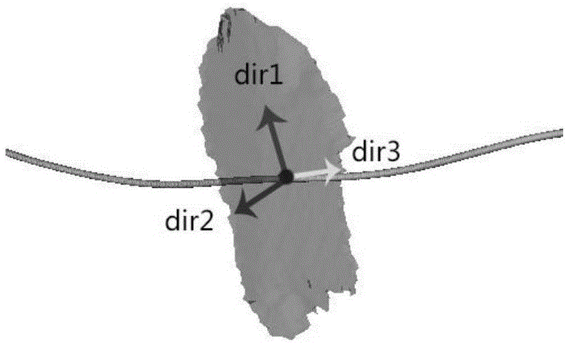
[0032] In one embodiment of the present invention, the cavity wall is the inner wall of the colon, and the cavity wall expansion method is the intestinal wall expansion method. In other alternative embodiments, the cavity wall can also be tubular organs such as blood vessel walls and trachea walls. the inner wall.

[0033] Postprocessing of colon CT images usually involves the following steps:

[0034] Obtain CT colon data: Scan the subject twice in the prone and supine positions to obtain CT colon data in DIC...


PUM


Abstract

The invention provides an organ cavity wall expanding method, comprising following steps: obtaining the mask and a centre line of an organ; initializing light direction of the points on the centre line, judging whether the mask has adhesion, if the mask has the adhesion, removing the adhesion; dividing the connected domain in the mask into a plurality of lamellas; solving the main directions namely a first direction, a second direction and a third direction of the lamellas in a three-dimensional coordinate system; solving the initial normal vector and the initial tangent vector of the points on the centre line; assigning the projection result of the initial normal vector in the plane in which the first direction and the second direction are located to the normal vector in the light direction, assigning the third direction or the reverse direction of the third direction to the tangent vector in the light direction; sampling the cavity wall according to the center line and the light direction obtained in the steps, mapping the sampling result to a two-dimensional plane to generate a cavity expanded two-dimensional view. The expanding effect of the cavity wall is improved through the settings.

Description

technical field [0001] The present invention relates to the processing of tomography (Computed Tomography, CT for short) images in the medical field, and in particular to a method for expanding cavity walls of tubular organs such as bronchi, blood vessels, and colon based on light projection. Background technique [0002] The colon is an important luminal organ of the human body. Since its birth in 1994, Computed Tomography Colonography (CTC) has become a commonly used medical imaging technology for detecting colon diseases through continuous development. Computed tomography colonography (CTC) has a similar effect to colonoscopy and can image the entire colon with less invasiveness. In the post-processing of colon CT images, the expansion of the intestinal wall is to convert the 3D view of the colon lumen into a 2D plane view, so as to facilitate the observation and comparison of the internal tissues of the colon, find and display colonic lesion tissues, and facilitate subse...

Claims


Application Information

Patent Timeline
Patent Type & Authority Applications(China)
IPC IPC(8): G06T3/00
CPCG06T3/0037
Inventor 孟晓林翁馨
Owner SHANGHAI UNITED IMAGING HEALTHCARE
Why Eureka
Try Eureka