Positioning method and apparatus for the liver scope in medical image

A medical image and positioning method technology, applied in the field of medical image processing, to achieve the effect of avoiding complex calculations and uncertain factors

Active Publication Date: 2016-06-15
SHANGHAI UNITED IMAGING HEALTHCARE
View PDF3 Cites 21 Cited by
  • Summary
  • Abstract
  • Description
  • Claims
  • Application Information

AI Technical Summary

Problems solved by technology

[0006] The problem to be solved by the present invention is to provide a method and device for locating the liver range in medical images, which can solve the above-mentioned problems in the existing liver locating methods, and quickly and accurately locate the liver range in the body data

Method used


Image

  • Positioning method and apparatus for the liver scope in medical image
  • Positioning method and apparatus for the liver scope in medical image
  • Positioning method and apparatus for the liver scope in medical image

Examples


Embodiment Construction

[0026] In order to make the above objects, features and advantages of the present invention more comprehensible, specific implementations of the present invention will be described in detail below in conjunction with the accompanying drawings. In the following description, specific details are set forth in order to provide a thorough understanding of the present invention. However, the present invention can be implemented in many other ways than those described here, and those skilled in the art can make similar extensions without departing from the connotation of the present invention. Accordingly, the present invention is not limited to the specific embodiments disclosed below.

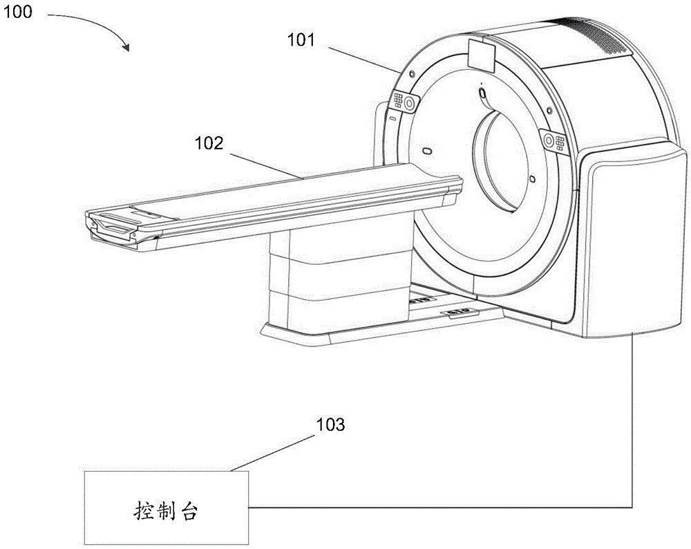
[0027] figure 1 It is a structural diagram of a medical imaging device, and a computed tomography device (CT, Computed Tomography) is taken as an example for illustration. see figure 1 , the computed tomography device 100 generally includes three parts: a frame 101 , a scanning table 102 and a co...


PUM


Abstract

The invention discloses a positioning method and apparatus for the liver scope in a medical image. The positioning method comprises the steps: acquiring a three dimensional medical image which is formed by a group of two dimensional medical images and covers the liver; determining the position the rib, in the three dimensional medical image, in the two dimensional medical images; based on the position of the rib, determining the position of the bottom of the liver in the two dimensional medical images; determining the position of the top of the liver in the two dimensional medical images; and according to the positions of the bottom and the top of the liver in the two dimensional medical images, determining the scope of the liver in the three dimensional medical image. The positioning method and apparatus for the liver scope in a medical image only need the current image data and have no demand for the prior information of a training sample or a probability graph.

Description

technical field [0001] The invention relates to the field of medical image processing, in particular to a method and device for locating a liver range in a medical image. Background technique [0002] In large-scale medical imaging equipment, it usually involves image processing for different organs, computer-aided diagnosis or quantitative analysis of index parameters. For this kind of medical image data, the image volume data (volumedata) covering the area of ​​the organ to be analyzed is generally collected and reconstructed when the medical imaging equipment is scanned, and then the application module corresponding to the organ to be analyzed performs specific diagnostic analysis. The image volume data here is a 3D medical image, usually composed of a set of 2D medical graphics. For example, image volume data obtained by image acquisition of a human torso can be used for diagnostic analysis of multiple organs (such as lungs, heart, liver, or intestinal tract). In each ...

Claims


Application Information

Patent Timeline
Patent Type & Authority Applications(China)
IPC IPC(8): G06T7/00G06T7/70
CPCG06T2207/30056
Inventor 吴柯韩妙飞
Owner SHANGHAI UNITED IMAGING HEALTHCARE
Why Eureka
Try Eureka