A Medical Image Segmentation Method Based on Statistical Deformation Model

A deformation model and medical image technology, applied in the field of medical imaging, can solve problems such as high complexity, low efficiency, and heavy workload of organ image segmentation, and achieve the effect of avoiding segmentation errors

Active Publication Date: 2018-11-16
NORTHWEST UNIV
View PDF5 Cites 0 Cited by
  • Summary
  • Abstract
  • Description
  • Claims
  • Application Information

AI Technical Summary

Problems solved by technology

[0006] In order to solve the problems of large workload, high complexity and low efficiency of organ image segmentation, the present invention proposes an organ-assisted localization and segmentation method based on statistical deformation model

Method used


Image

  • A Medical Image Segmentation Method Based on Statistical Deformation Model
  • A Medical Image Segmentation Method Based on Statistical Deformation Model
  • A Medical Image Segmentation Method Based on Statistical Deformation Model

Examples


Embodiment Construction

[0053] The present invention will be further described in detail below in conjunction with the accompanying drawings. It should be noted that the described embodiments are only intended to facilitate the understanding of the present invention, rather than limiting it in any way.

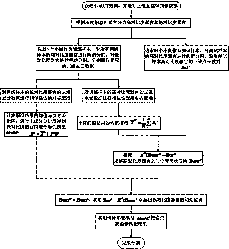
[0054] The present invention will be further described below in conjunction with accompanying drawing:

[0055] Step 1: Obtain mouse CT tomographic data:

[0056] Fix the experimental mice injected with the contrast agent on the imaging table of the Micro-CT imaging system, adjust the positions of the X-ray tube, the rotating table and the X-ray flat panel detector so that the centers of the three are in a straight line, and perform 360-degree imaging on the mice. High-degree irradiation scanning, acquisition of projection data, and three-dimensional reconstruction of projection data by filter back projection method to obtain mouse CT tomographic data.

[0057] The mouse CT volume data used in the e...


PUM


Abstract

The present invention discloses an organ-assisted positioning and segmentation method based on a statistical deformation model, which includes the following steps: (1) Obtaining biological CT data; (2) Dividing high- and low-contrast organs, selecting training samples in the CT data to extract corresponding Statistical prior information; (3) Establish a statistical deformation model of low-contrast organs; (4) Based on the correlation between high- and low-contrast organs, use high-contrast organs to assist in locating the initial position of low-contrast organs; (5) Assist Based on the positioning, the organs are searched and segmented along the normal direction of the marked points. The organ segmentation method used in the present invention can quickly and automatically find the initialization position of the organ, use the statistical deformation model to fuse prior information such as the position and shape of the tissue and organ, and quickly and systematically complete the segmentation of the organ, which greatly improves the efficiency of image segmentation. An efficient method for organ segmentation.

Description

technical field [0001] The invention belongs to the field of medical imaging, and relates to a medical image segmentation method based on a statistical deformation model. Background technique [0002] Medical image segmentation plays an extremely important role in modern medical research, clinical diagnosis, pathological analysis and treatment. It is a prerequisite for biomedical image analysis and a necessary step for human beings to understand the structure and function of tissues and organs. However, due to the diversity and complexity of human organs, the segmentation problem has always been a hot issue for researchers to explore. [0003] Commonly used methods in image segmentation include: edge detection method, threshold segmentation method, region growing method, active contour model method, etc. However, these methods have their own limitations: the edge detection method describes the process of grayscale change in the image based on the physical process that cause...

Claims


Application Information

Patent Timeline
Patent Type & Authority Patents(China)
IPC IPC(8): G06T7/10G06T7/60
CPCG06T7/60G06T2207/10004G06T2207/30004
Inventor 侯榆青王宇慧赵凤军贺小伟郭红波高培
Owner NORTHWEST UNIV
Why Eureka
Try Eureka