
Scanned image reestablishment method and device

A technology for scanning images and images, applied in the medical field, can solve the problems of subject suffering, main image not meeting requirements, inappropriate, etc.

Active Publication Date: 2016-08-31
NEUSOFT MEDICAL SYST CO LTD
View PDF8 Cites 6 Cited by
  • Summary
  • Abstract
  • Description
  • Claims
  • Application Information

AI Technical Summary

Problems solved by technology

[0003] Medical personnel set the main scanning conditions according to their own experience, but due to subjective or objective factors, the main scanning conditions set by medical personnel may not be suitable. When the medical equipment uses the main scanning conditions to perform main scanning on the subject, the obtained main image May not meet the requirements, for example, the image quality of the main image cannot meet the diagnostic requirements, resulting in the inability to use the currently obtained main image to diagnose the subject
[0004] In this case, the medical staff need to reset the main scanning conditions, and the medical equipment re-scans the subject under the new main scanning conditions, resulting in the subject being subjected to additional scanning radiation

Method used


Image

  • Scanned image reestablishment method and device
  • Scanned image reestablishment method and device
  • Scanned image reestablishment method and device

Examples


Embodiment Construction

[0022] The terminology used in this application is for the purpose of describing specific embodiments only, not to limit the application. As used in this application and the claims, the singular forms "a", "the" and "the" are intended to include the plural forms as well, unless the context clearly dictates otherwise. It should also be understood that the term "and / or" as used herein is meant to include any and all possible combinations of one or more of the associated listed items.

[0023] It should be understood that although the terms first, second, third, etc. may be used in this application to describe various information, the information should not be limited to these terms. These terms are only used to distinguish information of the same type from one another. For example, without departing from the scope of the present application, first information may also be called second information, and similarly, second information may also be called first information. Dependin...


PUM


Abstract

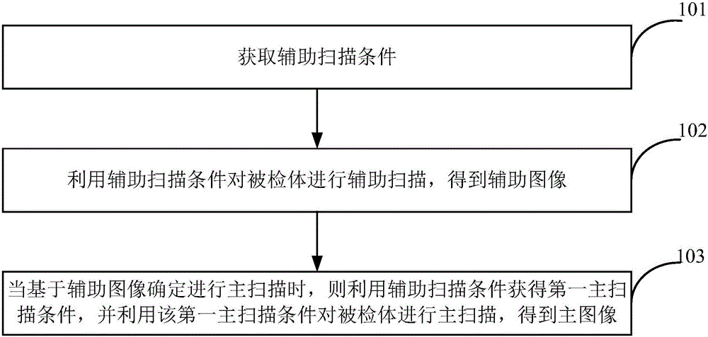
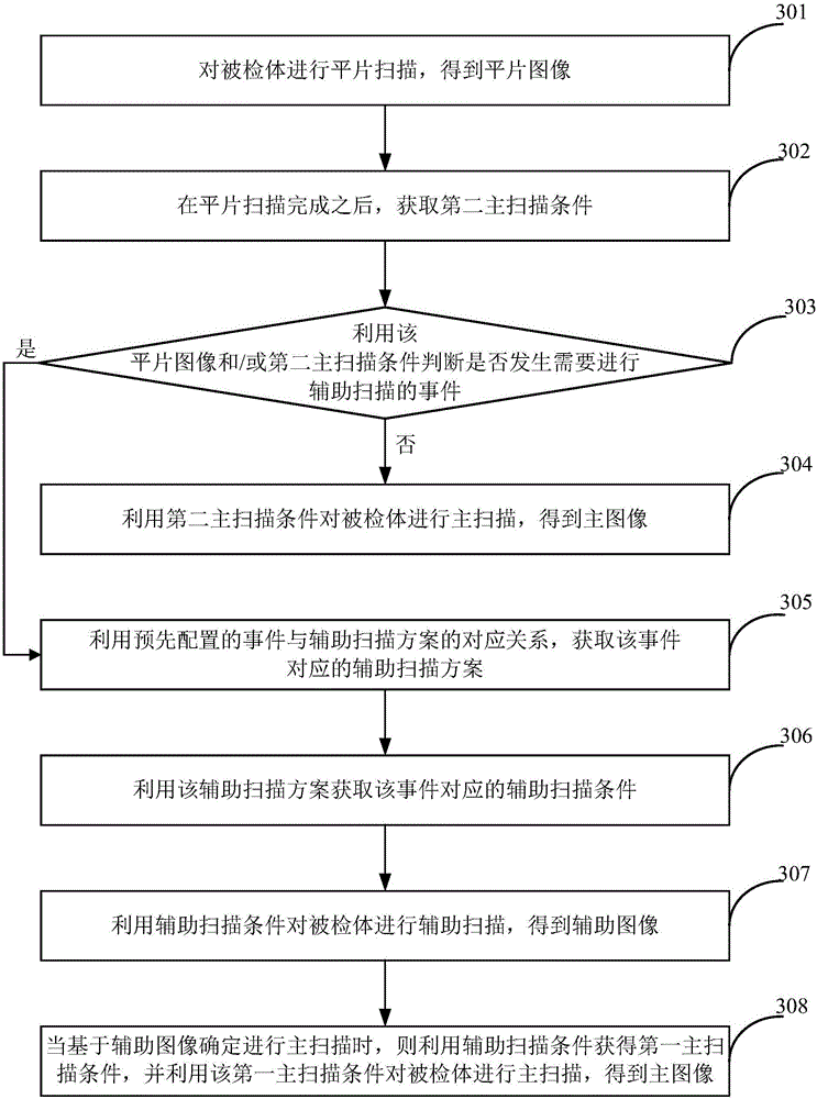
The invention provides a scanned image reestablishment method and device. The method includes the steps that an auxiliary scanning condition is obtained; the auxiliary scanning condition is utilized to conduct auxiliary scanning on an examined body, and an auxiliary image is obtained; when it is determined that main scanning is conducted based on the auxiliary image, a first main scanning condition is obtained through the auxiliary scanning condition, the first main scanning condition is utilized to conduct main scanning on the examined body, and a main image is obtained. By means of the scanned image reestablishment method and device, auxiliary scanning is added between plain film scanning and main scanning, quality of the main image can meet the diagnosis requirement, therefore main scanning conducted on the examined body repeatedly can be avoided, and extra scanning radiation conducted on the examined body is avoided.

Description

technical field [0001] The present application relates to the field of medical technology, in particular to a scanning image reconstruction method and device. Background technique [0002] For medical equipment that collects medical images, for example, common CT (Computed Tomography, electronic computer tomography) equipment, in order to collect images that can be used for diagnosis (for the convenience of description, it is referred to as the main image herein), conventional The method is: first scan the subject on a plain film to obtain a plain film image, and determine the scanning range and scanning parameters of the subject through the plain film image. After the plain film scanning is completed, the subject is scanned based on the scanning conditions set by the medical staff to obtain the main image. For convenience of description, the scanning process for obtaining the main image is referred to as main scanning, and the corresponding scanning conditions are referred...

Claims


Application Information

Patent Timeline
Patent Type & Authority Applications(China)
IPC IPC(8): A61B6/03G06T7/00
CPCA61B6/032A61B6/52G06T7/0012G06T2207/30004G06T2207/10104G06T2207/10088G06T2207/10081A61B6/488A61B6/545G06T11/003A61B6/5205
Inventor 王洪波
Owner NEUSOFT MEDICAL SYST CO LTD
Learn More