

G enterovirus detection antibody double sandwich ELISA detection kit
A technology of enterovirus and enzyme-labeled antibody, applied in the biological field, can solve the problems of lack of research on virus detection and diagnosis methods and its prevention and control, and achieve the effects of not affecting the normal production of animals, improving sensitivity and specificity, and being convenient to use
- Summary
- Abstract
- Description
- Claims
- Application Information
AI Technical Summary
Problems solved by technology
Method used
Image

Examples
Embodiment 1
[0030] Example 1 Construction of recombinant plasmid for prokaryotic expression of G type enterovirus structural protein VP1
[0031] 1. Primer design
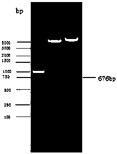
[0032] According to the base composition of the target sequence, design primers for amplification VP1 The upstream and downstream of the gene contain EcoRI and XhoI restriction sites respectively. The primer sequences are as follows:
[0033] Upstream primer: 5' TT GAATTC CAGGCTGGGTATGTGACT 3' (EcoRI)
[0034] Downstream primer: 5'T CTCGAG TTAATGAAAATCACTGAGGGGTT 3' (XhoI)
[0035] 2. Gene amplification
[0036] Genomic RNA of G enteroviruses was extracted by the conventional Trizol method, and cDNA was obtained by using a commercially available conventional reverse transcription kit according to the instructions, and amplified using it as a template VP1 Gene, PCR reaction system:
[0037]
[0038] PCR operating conditions: pre-denaturation at 94°C for 3 min; denaturation at 94°C for 1.5 min, annealing at 54°C for...
Embodiment 2
[0041] Example 2 Expression and purification of VP1 recombinant protein
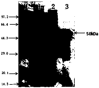
[0042]After the identified positive recombinant plasmid pGEX-4T-1-VP1 was transformed into Escherichia coli BL21(DE3) competent cells, plated and incubated overnight at 37°C, a single colony was picked and inoculated into 3 mL of 100 µg / mL ampicillin containing Cultivate overnight in LB liquid medium, then inoculate 1 mL of the above culture into 200 mL of LB liquid medium containing 100 μg / mL ampicillin and culture at 37 °C until logarithmic growth phase (OD 600 =0.6), adding IPTG (final concentration 1 mmol / L), inducing culture at 20°C for 3 h, and detecting by SDS-PAGE, the recombinant target protein VP1 was obtained, as figure 2 Shown (lane 2). Centrifuge the bacterial cells, and after ultrasonic disruption, wash with 1.5M urea for 3 times to obtain high-purity recombinant protein, such as figure 2 Shown (lane 3).
Embodiment 3
[0043] Example 3 Preparation of Anti-Sheep Enterovirus VP1 Recombinant Protein Monoclonal Antibody and Polyclonal Antibody
[0044] 1. Preparation of anti-VP1 monoclonal antibody: select 6-8 week-old healthy BALB / c mice to emulsify and purify VP1 recombinant protein with complete Freund's adjuvant, and inject 100 μg subcutaneously into each mouse at multiple points, and then every 14 days Boost the immunization once in the same way, and collect serum to test the titer at the same time. For the last booster immunization, 100 μg of purified protein was directly injected intraperitoneally. After 3 to 5 days, the splenocytes of the immunized mice were mixed with SP2 / 0 in a fusion tube, centrifuged at 300 g for 10 min, the supernatant was discarded, and the cells were shaken to make the two Mix the seeded cells as evenly as possible, then slowly add the preheated PEG-4000 solution dropwise within 60 sec, and then slowly add serum-free 1640 medium to terminate the fusion, let it sta...
PUM
 Abstract
Description
Claims
Application Information