Manufacturing of glaucoma internal drainage replacement bionic support and application method thereof

A glaucoma and internal drainage technology, applied in wound drainage, ophthalmic surgery, medical science, etc., can solve the problems of high requirements for surgeons, complicated operations, and high cost, and achieve the effect of solving the scarring of the orifice

Active Publication Date: 2016-10-12
苏州朗目医疗科技有限公司
View PDF9 Cites 13 Cited by
  • Summary
  • Abstract
  • Description
  • Claims
  • Application Information

AI Technical Summary

Problems solved by technology

However, due to the need for special equipment and materials, the dilatation of the viscous tubules is expensive, the operation is complicated, and the requirements for the surgeon are high, and the current research shows that the dilatation of the viscous tubules is only suitable for open-angle glaucoma, and the angle-closure glaucoma is due to the angle The iris is blocked by adhesions, making it a contraindication to surgery and cannot be widely used clinically

Method used


Image

  • Manufacturing of glaucoma internal drainage replacement bionic support and application method thereof
  • Manufacturing of glaucoma internal drainage replacement bionic support and application method thereof
  • Manufacturing of glaucoma internal drainage replacement bionic support and application method thereof

Examples


Embodiment 1

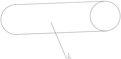
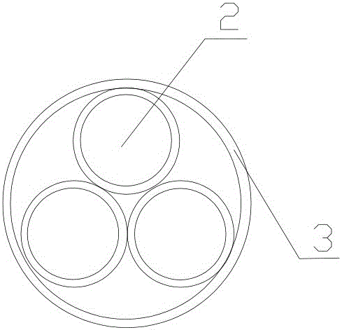
[0021] A glaucoma internal drainage substitute bionic bracket, comprising a cylindrical tube body 1, the middle of the tube body 1 is a hollow structure, and three straight tubes 2 are arranged in the hollow structure in the middle of the tube body 1, and the straight tube body 1 The tube 2 supports the tube wall 3 of the tube body 1 , the straight tube 2 is a round hole, and the three circular straight tubes 2 are arranged in a triangle in the hollow structure of the tube body 1 . The tube body 1 has a tube length of 6-10 mm and a cross-sectional diameter of 300 μm. The bionic bracket is made of polyurethane.

Embodiment 2

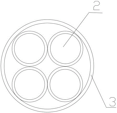
[0023] A kind of glaucoma internal drainage instead of bionic stent, comprising a cylindrical tube body 1, the middle of the tube body 1 is a hollow structure, and four straight tubes 2 are arranged in the hollow structure in the middle of the tube body 1, the described The straight pipe 2 supports the pipe wall 3 of the pipe body 1, the straight pipe 2 is a round hole, and the four circular straight pipes 2 are arranged in the hollow structure of the pipe body 1 in a quadrangular shape. The tube body 1 has a tube length of 6 mm and a cross-sectional diameter of 300 μm. The bionic bracket is made of polyurethane.

Embodiment 3

[0025] A kind of glaucoma internal drainage instead of a bionic stent, comprising a cylindrical tube body 1 with a hollow structure in the middle of the tube body 1, and six straight tubes 2 are arranged in the hollow structure in the middle of the tube body 1, the described The straight pipe 2 supports the pipe wall 3 of the pipe body 1 , the straight pipe 2 is polygonal, and the several polygonal straight pipes 2 are closely arranged on the inner wall of the hollow structure of the pipe body 1 . The tube body 1 has a tube length of 6 mm and a cross-sectional diameter of 300 μm. The bionic bracket is made of polyurethane.

[0026] Bionic stent placement process:

[0027] Routinely disinfect and spread drape on the operated eye, place a lid speculum, rinse the conjunctival sac with diluted iodophor solution, take 0.4ml 2% lidocaine in the subconjunctival local anesthesia, do superior rectus muscle traction suture fixation, press the dial of the clock Direction meter, incis...


PUM


Abstract

Provided are manufacturing of a glaucoma internal drainage replacement bionic support and an application method thereof. Through proper placing and fixation, aqueous fluid smoothly flows into a Schlemm tube through a pipe orifice supported by the bionic support, through filling of aqueous fluid, the collapse Schlemm tube is expanded again, and the aqueous fluid is drained to a collecting tube and an aqueous vein, to achieve an objective of reducing intraocular tension and treating glaucoma. A new glaucoma internal drainage replacement bionic support implantation greatly reduces operation cost and reduces operation cost for a patient while maintaining advantages of a nonpenetrating glaucoma surgery. Combined with a trabeculectomy, adaptation diseases of the glaucoma internal drainage replacement bionic support are expanded, and new thinking and suggestion are provided for treatment of glaucoma.

Description

technical field [0001] The invention relates to the field of medical equipment, in particular to a method for preparing and using a glaucoma internal drainage replacement bionic bracket. Background technique [0002] Glaucoma is a kind of optic nerve damage disease caused by pathological high intraocular pressure. Controlling intraocular pressure has become the main means of glaucoma treatment. Although anti-glaucoma drugs and laser technology provide the technology and possibility to control intraocular pressure, due to the particularity of the disease, a considerable number of glaucoma patients have to undergo surgery. At present, the mainstream operation method for glaucoma is trabeculectomy, but glaucoma filtration surgery has many uncertain factors and relatively high complications, such as postoperative low intraocular pressure, shallow anterior chamber, choroidal detachment, cystoid macular edema and Surgery failure, filtering bleb scarring and other problems. Seeki...

Claims


Application Information

Patent Timeline
Patent Type & Authority Applications(China)
IPC IPC(8): A61F9/007
CPCA61F9/00781A61F2230/0006A61F2230/0017A61F2240/001A61M25/0026A61M27/002A61F9/007
Inventor 梁远波李星熠石长灿胡城
Owner 苏州朗目医疗科技有限公司
Why Eureka
Try Eureka