Capsule endoscope image data redundancy removing method

A capsule endoscope and image data technology, applied in the field of image processing, can solve problems such as unfavorable use and large amount of calculation, and achieve the effects of improving diagnostic efficiency and accuracy, improving accuracy and reducing the total number of pictures

Inactive Publication Date: 2016-10-26
安翰科技(武汉)股份有限公司
View PDF4 Cites 21 Cited by
  • Summary
  • Abstract
  • Description
  • Claims
  • Application Information

AI Technical Summary

Problems solved by technology

This technology adopts the image registration technology, which considers the global motion model and the local motion model

Method used


Image

  • Capsule endoscope image data redundancy removing method
  • Capsule endoscope image data redundancy removing method
  • Capsule endoscope image data redundancy removing method

Examples


Embodiment Construction

[0019] The present invention will be further described in detail below in conjunction with the accompanying drawings and specific embodiments.

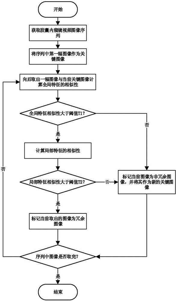
[0020] Such as Figure 1~3 Described capsule endoscope image data de-redundancy method, it comprises the steps:

[0021] Step 1: After the patient swallows the capsule endoscope, the capsule endoscope collects image data in the digestive tract, and the image sequence in the collected image data has M images (color images) arranged according to the shooting time. The image corresponding to the first shooting moment is used as the key image, and enters step 2;

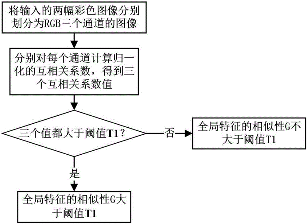
[0022] Step 2: Take the image at the next moment adjacent to the key image as the comparison image, and calculate the global feature similarity between the key image and the comparison image, that is, extract the three components of R, G, and B of the key image and compare The R, G, B three components of the image, respectively calculate the normalized cross-correlation coeffi...


PUM


Abstract

The invention discloses a capsule endoscope image data redundancy removing method comprising the steps as follows: (1) taking a first image in an image sequence as a key image; (2) taking an image next to the key image as a contrast image, and calculating the global characteristic similarity between the key image and the contrast image; (3) judging whether normalized cross-correlation coefficients are all greater than a threshold, if the normalized cross-correlation coefficients are all greater than the threshold, determining that the global characteristic similarity is greater than a threshold, further calculating the local characteristic similarity between the key image and the contrast image, and going to step (4), or, taking the current contrast image as a new key image and returning to step (2); and (4) judging whether the local characteristic similarity between the key image and the contrast image is greater than a preset threshold, if the local characteristic similarity between the key image and the contrast image is greater than the preset threshold, marking the current contrast image as a redundant image and deleting the contrast image, or, taking the current contrast image as a new key image and returning to step (2). Through the method, the total number of images needing to be checked by doctors can be reduced, and the diagnosis efficiency and accuracy can be improved.

Description

technical field [0001] The invention relates to the technical field of image processing, in particular to a method for removing redundancy of image data of a capsule endoscope. Background technique [0002] The wireless capsule endoscope can inspect the entire digestive tract in a painless and non-invasive way, which is a revolutionary technological breakthrough. The patient swallows a capsule, the capsule advances along the digestive tract, and takes image data. The capsule endoscope needs to work for 6 to 8 hours in total and collect about 50,000 images. A large amount of image data makes the doctor's inspection work difficult and difficult. time consuming. Professional doctors usually need about 2 hours to complete the examination of the entire image sequence. Sometimes the abnormality of the digestive tract only exists in a few frames of images. In addition, there is usually a lot of redundant information in the images collected by the capsule endoscope. Doctors may mi...

Claims


Application Information

Patent Timeline
IPC IPC(8): G06T7/00
CPCG06T7/0012G06T2207/10068G06T2207/30096
Inventor 张行袁文金张皓王新宏段晓东肖国华
Owner 安翰科技(武汉)股份有限公司
Why Eureka
Try Eureka