18-Lead holographic dynamic and static electrocardiographic analysis method and system

A technology of lead system and analysis method, which is applied in the fields of electrical digital data processing, medical science, medical equipment, etc., and can solve the problems of time-consuming and laborious, inability to synchronize homologous sampling and synchronous analysis, etc.

Inactive Publication Date: 2016-11-09
BEIJING MEDSUN MEDICAL EQUIP
View PDF6 Cites 9 Cited by
  • Summary
  • Abstract
  • Description
  • Claims
  • Application Information

AI Technical Summary

Problems solved by technology

This method is not only time-consuming and labor-intensive, but also cannot synchronize homologous sampling and simultaneous analysis with the above-mentioned 12 leads

Method used


Image

  • 18-Lead holographic dynamic and static electrocardiographic analysis method and system
  • 18-Lead holographic dynamic and static electrocardiographic analysis method and system
  • 18-Lead holographic dynamic and static electrocardiographic analysis method and system

Examples


Embodiment Construction

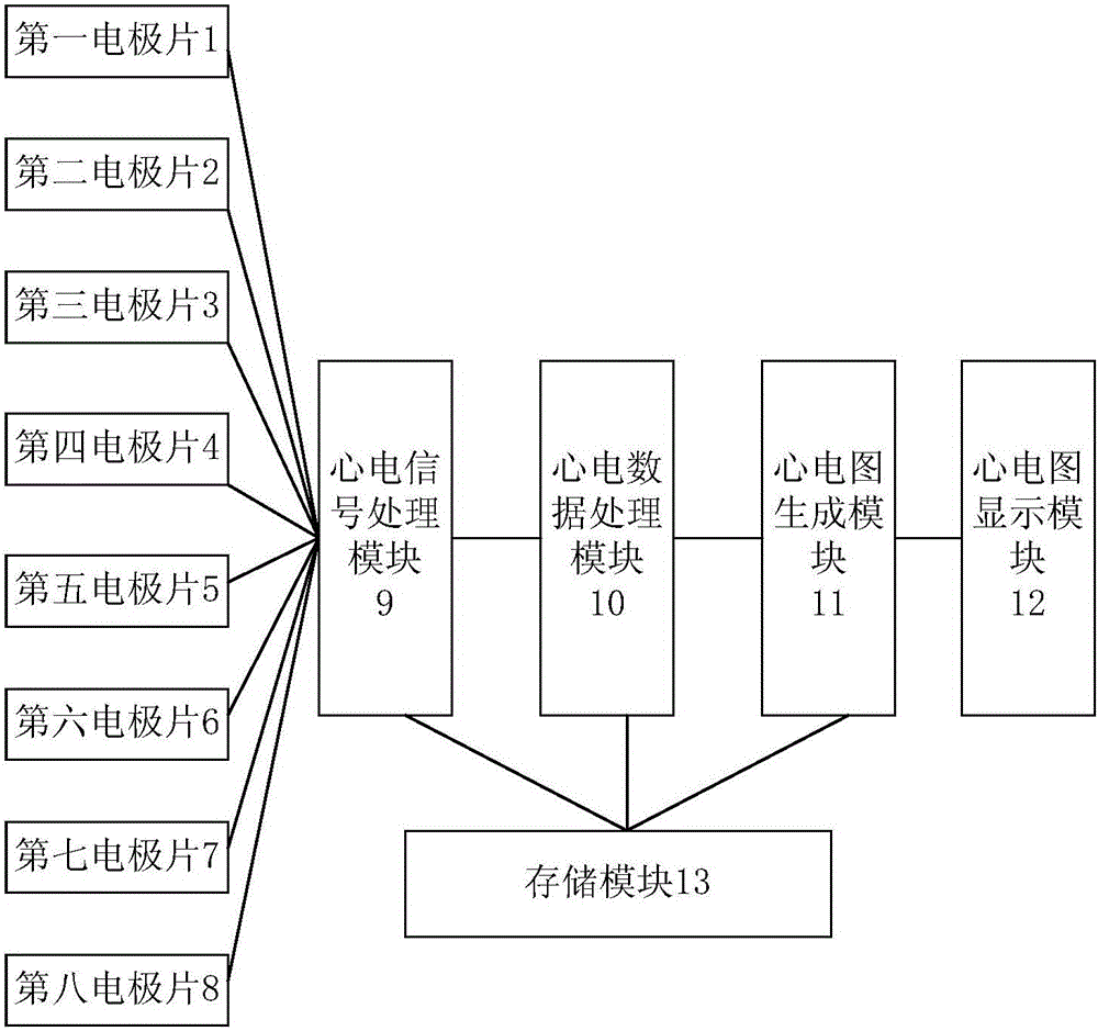
[0058] Embodiments of the present invention are described in detail below, and examples of the embodiments are shown in the drawings, wherein the same or similar reference numerals denote the same or similar elements or elements having the same or similar functions throughout. The embodiments described below by referring to the figures are exemplary and are intended to explain the present invention and should not be construed as limiting the present invention.

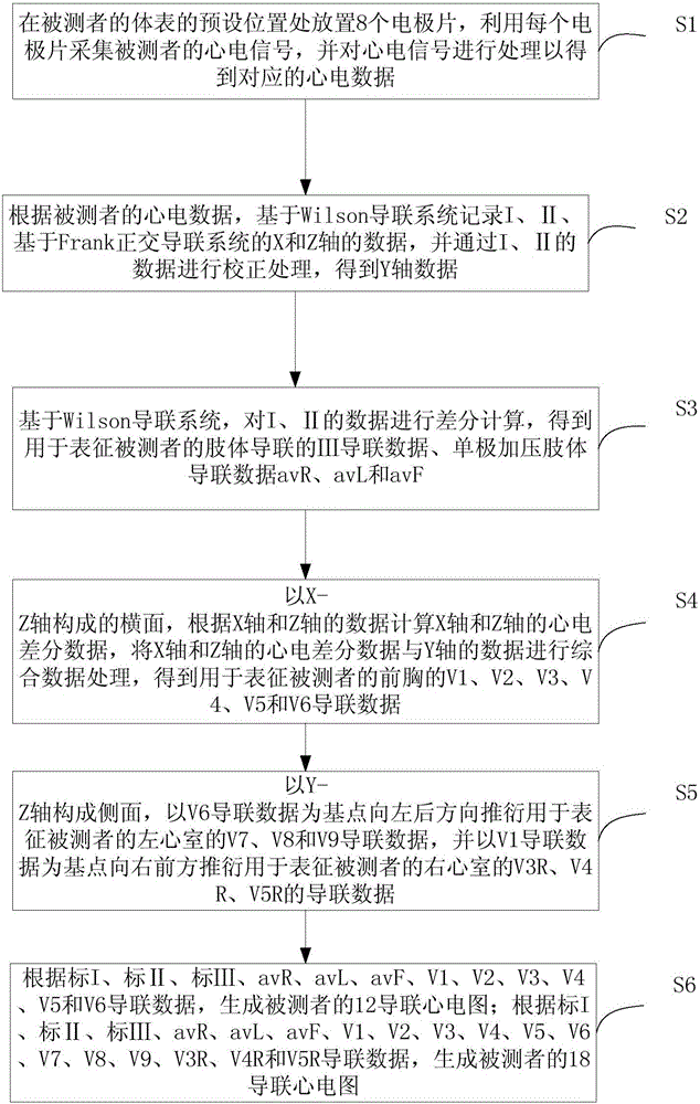
[0059] First, the theoretical basis of the 18-lead holographic dynamic and static electrocardiogram analysis method and system of the embodiment of the present invention will be described below. Theoretical basis of the present invention originates from electrocardiogram Einstein's triangle theory (galvanic couple-volume conduction theory), electrocardiographic vector diagram - electrocardiogram orthogonal electrocardiogram and electrocardiogram Figure II Basic theory of subprojection. ECG Figure II Subprojection t...


PUM


Abstract

The invention provides an 18-lead holographic dynamic and static electrocardiographic analysis method and system, comprising: placing 8 electrodes at preset positions on the body surface of a subject to collect electrocardiogram signals of the subject; based on Wilson lead system recordings I and II and X-axis and Z-axis data of Frank lead system, using I and II data and Y-axial data as basis; acquiring I, II, III, avR, avL and avF based on the Wilson lead system; acquiring V1-V6 lead data representative of the chest of the subject by using a cross section composed of X-Z axes; deriving V7-V9 lead data representative of the left ventricle of the subject by using Y-Z axes to form a side, and deriving to the front right, V3R-V5R lead data of the right ventricle of the subject by using V1 lead data as a basal point; generating 12 and 18 lead electrocardiograms of the subject. The invention implements the detection of 18-lead electrocardiograms based on the modified Wilson lead system and Frank orthogonal lead system.

Description

technical field [0001] The invention relates to the technical field of electrocardiogram monitoring, in particular to an 18-lead holographic dynamic and static electrocardiogram analysis method and system. Background technique [0002] The heart is a three-dimensional organ, and the electrocardiographic vector generated at each moment during the excitation process has a certain spatial position, that is, the three-dimensional relationship of up and down, left and right, and front and back reflects this three-dimensional vector, which is called a space vector. Because the ECG vector can display the phase relationship of the ECG, it makes up for some deficiencies in the diagnosis of the ECG. Both the ECG and the ECG vector are the main low-frequency components in the ECG information collected from the body surface, and they have a common theoretical basis. It's just that the lead system and the way of expression are different. [0003] In the conventional electrocardiogram in...

Claims


Application Information

Patent Timeline
Patent Type & Authority Applications(China)
IPC IPC(8): A61B5/0402A61B5/0408G06F19/00
CPCA61B5/282A61B5/316A61B5/318G16H40/40
Inventor 孙龙杜永盛
Owner BEIJING MEDSUN MEDICAL EQUIP
Why Eureka
Try Eureka