
A medium-term corneal preservation solution without serum components
A cornea and preservation solution technology, applied in the field of serum-free corneal mid-term preservation solution and its preparation, can solve the problems of preservation crisis, high price, inability to popularize and apply, etc.
- Summary
- Abstract
- Description
- Claims
- Application Information
AI Technical Summary
Problems solved by technology
Method used
Image

Examples
Embodiment 1
[0022] Preparation of corneal preservation solution:
[0023] 1. 500ml of compound electrolyte intraocular irrigation solution is used as the mother solution;
[0024] 2. Take 25g of chondroitin sulfate, add 275ml of deionized water, heat, dissolve and autoclave to make chondroitin sulfate solution; add 2.93g of sodium chloride, 0.6g of magnesium sulfate, 2.72g of potassium dihydrogen phosphate, add 100ml of water for injection to dissolve and autoclave , to make a sodium magnesium potassium salt solution; add 5 g of sodium hyaluronate, add 100 ml of water for injection to dissolve and sterilize by autoclaving, and make a sodium hyaluronate solution;
[0025] 3. Take a sterile container and add 500ml of compound electrolyte intraocular irrigation solution, 275ml of autoclaved chondroitin sulfate, 100ml of sodium magnesium potassium salt solution, and 100ml of sodium hyaluronate solution; add 3g of reduced glutathione and dexamethasone injection 1.5mg, levofloxacin injection 0...
Embodiment 2
[0031] Detection of the effect of corneal preservation solution:
[0032] 1. Get fresh pig eyeballs (obtained within 2 hours after slaughter), aseptically treat them, and cut out the whole cornea with 1-2mm sclera limbus, and place them in 20ml of the corneal preservation solution prepared in Example 1 and 20ml of Optisol as a contrast Corneal active preservation solution;
[0033] 2. Store at 4°C for 3 days, 7 days and 14 days for testing the preservation effect.
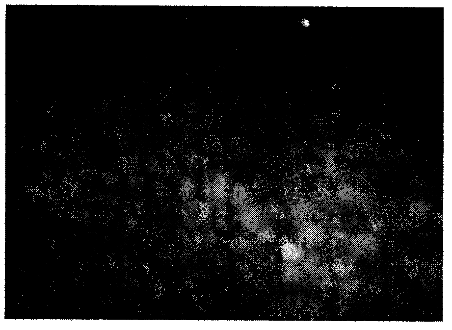
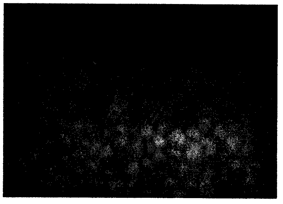
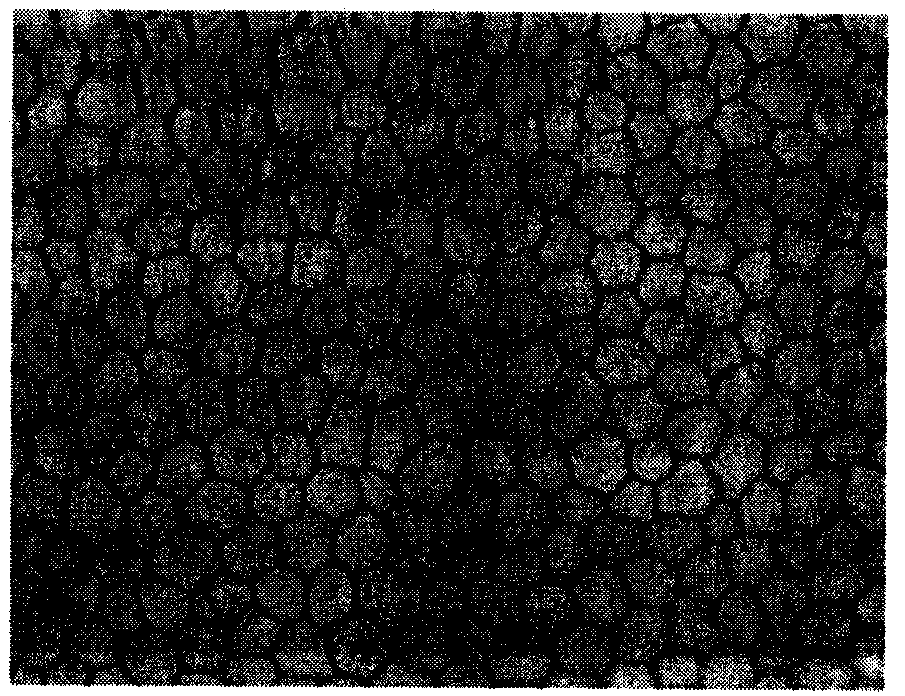
[0034] 3. After 3 days of storage, the corneal slices were taken out and stained with trypan blue. It was found that the number of corneal endothelial cells, the hexagonal shape and the transparency of the cornea preserved in the corneal active preservation solution were not different from those in the contrast Optisol corneal active preservation solution;
[0035] 4. After 1 week of storage, the corneal slices were taken out and stained with trypan blue. It was found that the number of corneal endothelial cells, ...
Embodiment 3
[0038] Corneal medium-term preservation solution to preserve cat cornea for penetrating keratoplasty:
[0039] 1. Take fresh (obtained within 2 hours after slaughter) cat corneal grafts in the corneal preservation solution prepared in Example 1, store them in a refrigerator at 4°C for 2 weeks, and wait for transplantation;
[0040] 2. Six healthy domestic cats were anesthetized. A 7.0mm-diameter trephine was routinely used to drill and cut the central cornea. After the cornea was cut, the cat corneal grafts that had been stored for 2 weeks were placed on the cutting pillow, and a 7.25mm-diameter trephine was used to cut the central cornea. Drill the implant from the endothelial surface, cover it on the cat corneal implant hole, suture intermittently with 10 / 0 nylon suture, and form the anterior chamber watertight;
[0041] 3. Observe the transparency of corneal grafts with slit lamp on 1 day, 3 days, 5 days, 7 days and 14 days after operation, and detect the endothelial number...
PUM
 Abstract
Description
Claims
Application Information