
System and method for detecting a medical condition in a subject

A condition, animal technology, applied in the field of systems and methods for the detection of medical conditions in a subject, capable of addressing non-specificity and other issues

Inactive Publication Date: 2016-11-23
BIOSENSE MEDICAL
View PDF5 Cites 1 Cited by
  • Summary
  • Abstract
  • Description
  • Claims
  • Application Information

AI Technical Summary

Problems solved by technology

However, while imaging is quite sensitive, it is also relatively nonspecific

Method used


Image

  • System and method for detecting a medical condition in a subject
  • System and method for detecting a medical condition in a subject

Examples


Embodiment

[0089] Materials and methods:

[0090] cell line:

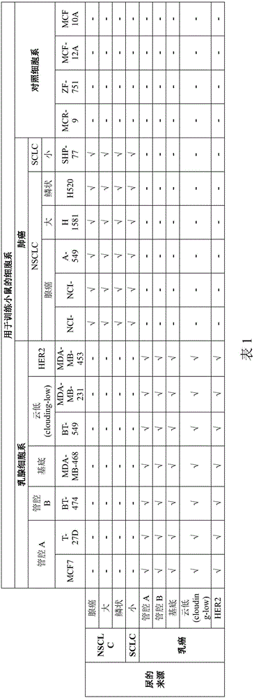
[0091] The following commercially available cell lines were used. Breast cancer cell lines: MCF7, T-47D, BT-474, MDA-MB-468, BT-549, MDA-MB-231, MDA-MB-453. Lung cancer cell lines: NCI-H1299, NCI-H2030, A-549, SHP-77, H1581 and H520. Control (healthy) cell lines: MCF-12A, MCF 10A MRC-9 and ZR-75-1.

[0092] tissue culture

[0093] at 5% CO 2 atmosphere, at 175cm 2 Cell lines were grown in RPMI 1640 medium or DMEM medium supplemented with 10% fetal bovine serum in cell culture flasks. Will be about 2x 10 6 cells were seeded in flasks and grown to approximately 95% confluency (7 x 10 6 cells), at which point a tissue culture is obtained.

[0094] tissue culture samples

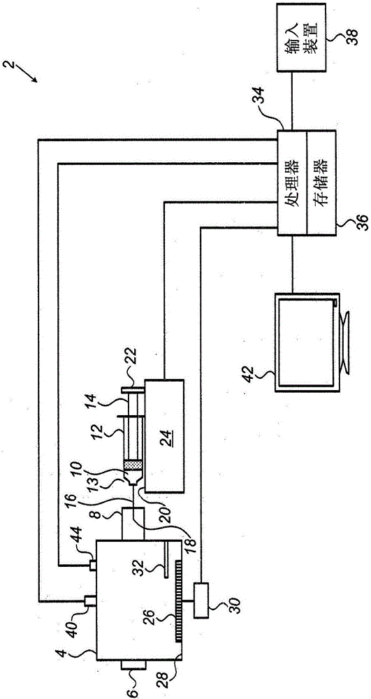
[0095] Attach silicon connectors to each culture flask. Tissue culture samples were obtained by piercing the silicon adapter using a 60 ml syringe with a 20G 1.5" needle and collecting the headspace of the culture in the syringe.

[0096] sa...


PUM


Abstract

A method for training an animal to detect a condition, such as cancer, in a human or animal individual is disclosed. A training sample is presented to the animal to be trained where, the training sample is a gaseous sample or vapor generated by a cell population associated with the predetermined condition. The population of cells associated with the predetermined condition may be, for example, a culture of an established cell line associated with the predetermined condition. Simultaneously with, or subsequent to, presentation of the training sample, the animal is subjected to an adverse stimulus, such as an electric shock. The animal is allowed animal to perform a first predetermined response in order to avoid, escape or terminate the adverse stimulus. The invention also provides a method for detecting a condition, such as cancer, in an individual in which a trained animal performs the predetermined response upon exposure to vapors from a body fluid of an individual affected with the condition.

Description

technical field [0001] The present invention relates to methods and systems for training animals to detect odors, and using trained animals to detect odors. Background technique [0002] The following prior art publications are considered relevant for understanding the background of the present invention: [0003] Sponring A, et al. (2009) Release of volatile organic compounds from thelung cancer cell line NCI-H2087 in vitro. Anticancer Res 29:419-426. [0004] Filipiak W, et al. Release of volatile organic compounds (VOCs) from thelung cancer cell line CALU-1 in vitro Cancer Cell International 2008,8:17. [0005] Matsumura et al., Urinary Volatile Compounds as Biomarkers for Lung Cancer: A Proof of Principle Study Using Odor Signatures in Mouse Models of Lung Cancer, Publication Date: January 27, 2010. [0006] Dragonieri, S., et al. An electronic nose distinguishes exhaled breath of patients with Malignant Pleural Mesothelioma from controls. Lung Cancer 75(3):326-31. J. ...

Claims


Application Information

Patent Timeline
IPC IPC(8): A01K1/03
CPCA01K1/031A01K15/02A01K15/029
Inventor 米哈尔·马克-达尼埃利阿舍·卡斯铁尔
Owner BIOSENSE MEDICAL
Learn More