Method and device for monitoring loss of body fluid and dislodgment of medical instrument from body

a technology of body fluid and monitoring device, which is applied in the direction of diagnostic recording/measuring, separation processes, applications, etc., can solve the problems of hemodialysis treatment, unintentional hazardous situation, and great pain

Inactive Publication Date: 2005-02-17
BRADLEY JON MOLL RODNEY L MOLL & ANN E MOLL FAMILY TRUST
View PDF15 Cites 385 Cited by
  • Summary
  • Abstract
  • Description
  • Claims
  • Application Information

AI Technical Summary

Benefits of technology

[0018] The present invention involves a device for monitoring loss of body fluid and dislodgment of a medical instrument from an access site of the body. The device involves a system that is designed for use as a critical medical monitor, provides the requisite electrical isolation, and provides an unobstructed view of the access site. The device incorporates a sterile and disposable sensor that fails-to-safe such that a loss of protection results in an alarm, can be reset in the case of small leaks, can be tested “in-situ”, without extra test equipment, alarms if the patch is removed or damaged in use, can stand alone and is not be required to be integrated into the alarm circuits of common, existing hemodialysis units, monitors the placement of the needle relative to its insertion point, has a monitored power supply, and alarms in the intentional removal of needle by patient.

Problems solved by technology

However, due to differences in protocols, techniques, or varying patient needs, some hemodialysis treatment may last six hours or even overnight.
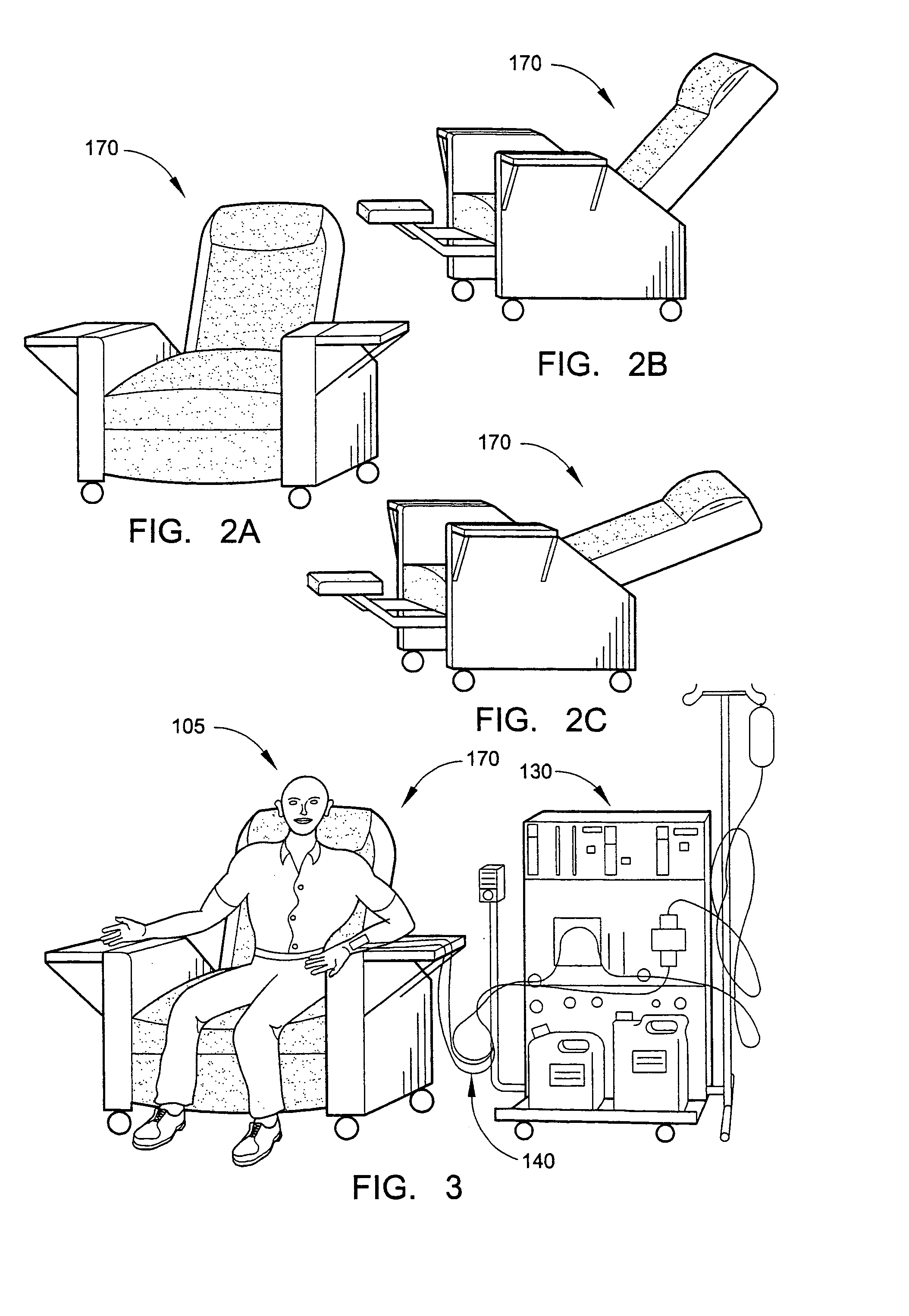
Some examples of how this hazardous situation may occur unintentionally include the bloodlines getting caught on the treatment chair during a positional change such as reclining from a seated position or siting up from a reclined position.
It may even happen when the tape on a patient simply comes off, due to dry or sweaty skin, and the needles slip out.
Other patients move around frequently and the many little tugs on the bloodline and tapings, and the constant pulling eventually loosens that tapings to the point that they come off and the fistula needle falls out.
Should the venous needle become partially dislodged during a treatment, the patients' blood may infiltrate (into the surrounding tissue areas), usually causing great pain, or it may leak out around the needle entry site, or a combination of both.
Obviously, even the most observant and dedicated of medical staff could not possibly watch each patient all of the time.
Significantly, the VPM will often fail to register a sufficient pressure change (to set off an alarm) due to the inherent “back pressure” developed in a venous blood line by the resistance of the viscous blood traveling through the relatively small orifice of the fistula needle.
This solution has limitations in that it is rather slow to respond as it is dependent on the reaction of the body to the problem.
Additionally, it fails to address the present concern of dislodgment.
While an infiltration is painful, may require surgery to correct and could even result in the loss of the limb, it is not immediately life threatening.
This method has not experienced commercial success, or widespread utilization.
It is believed this may be because the pressures illustrated appear to be on a very still patient, which is not a realistic assumption throughout a four-hour or longer treatment.
This arrangement does not adequately protect a patient as it could have a sensor failure or disconnect without alerting the staff of the sensor failure or disconnection.
Additionally, the device does not explicitly provide for compliance with the nonisolated patient connection requirements of Safe Current Limits for Eletromedical Apparatus as required by Applicable Document 2.3.
Daneshvar's unit also is not compliant with the nonisolated patient connection requirements for electrically sensitive patients.
Additionally, whether Daneshvar's unit will alarm depends on the absorbency of the gauze, which may be compromised due to being saturated by non-conductive fluids or by compression, or coated by certain medical gels, pastes or ointments.
However, neither of these allow for an unobstructed view of the access site.
Another problem they share is that they are designed for use as an integral part of a dialysis machine.
As such, they are specifically not designed for stand-alone use.
Of greater concern is the failure of any of these devices to fail in a safe manner.
If the unit fails for some reason, such as a dead battery, the protection is lost and the staff is not aware of it.
With some of the newer implanted tubing and other new types of vascular accesses, there is very little bleeding when a needle is removed, and, hence, limited opportunity for success in alerting the staff in the case of a rapid needle withdrawal, such as when a bloodline is caught by a passing foot, or when a mentally unstable patient intentionally removes the needle.
Additionally, in applications other than dialysis, the underlying region or substrates may not have a sufficient positive relative pressure to force out blood, body fluid or liquid to wet the sensor.
The reusable sensors described in the references mentioned above and elsewhere in the prior art involve the myriad of problems and costs associated with reusing soiled medical equipment including, but not by way of limitation, sterilization, clean storage, verification of the absence of the sterilant prior to use, reused devices being reused only by the original patient, quality problems due to subjective assessments, etc.

Method used


Image

  • Method and device for monitoring loss of body fluid and dislodgment of medical instrument from body
  • Method and device for monitoring loss of body fluid and dislodgment of medical instrument from body
  • Method and device for monitoring loss of body fluid and dislodgment of medical instrument from body

Examples


Embodiment Construction

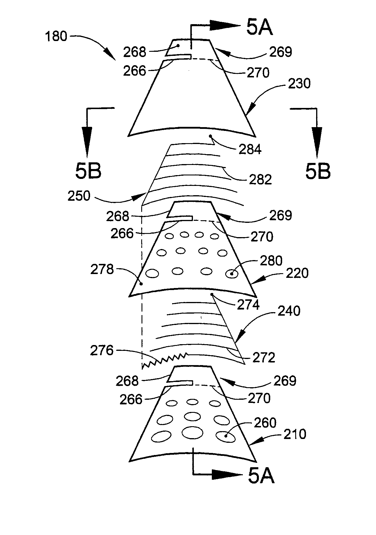
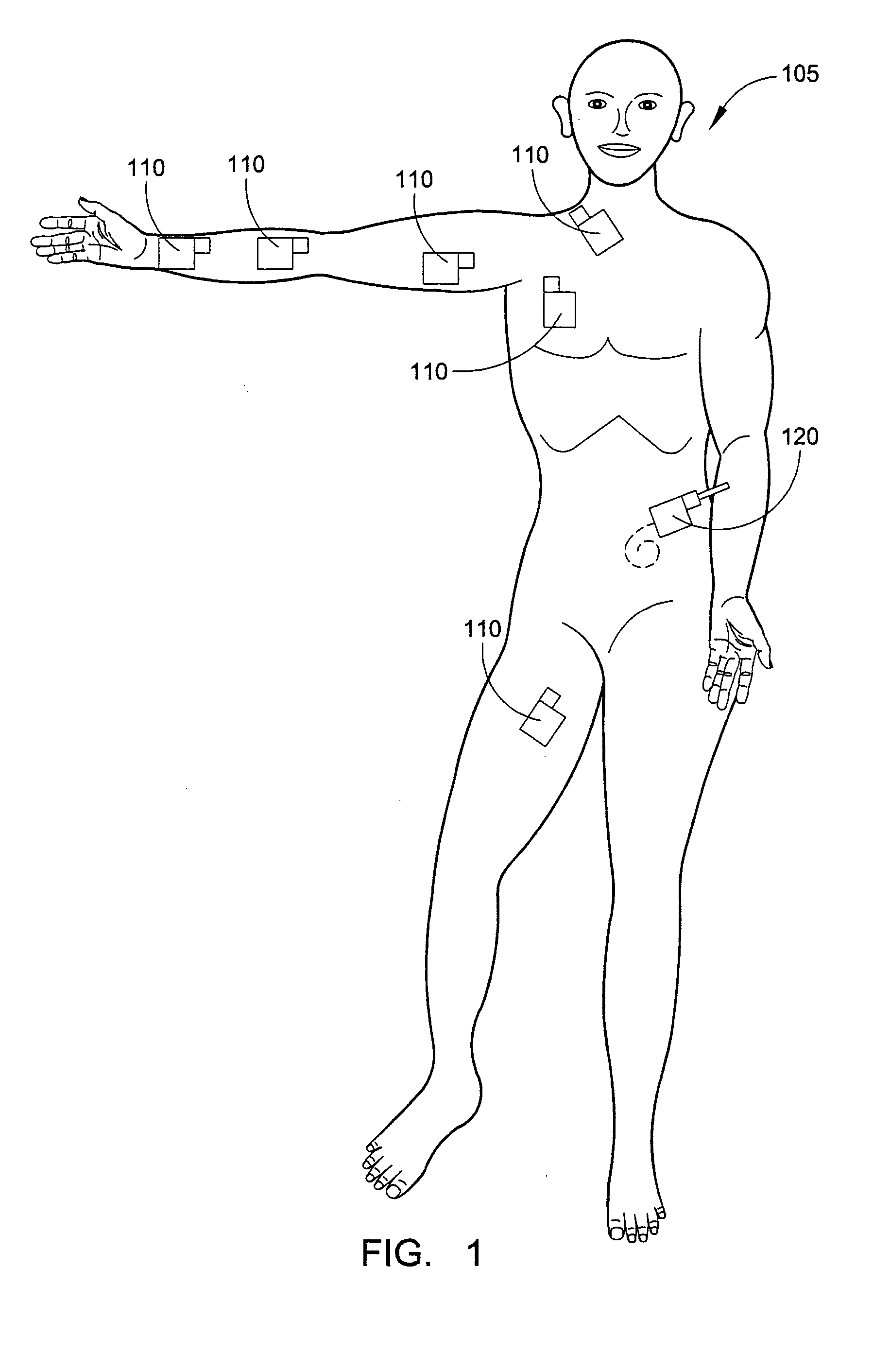
[0036] With reference to FIGS. 1-10, an embodiment of a system and method for monitoring a vascular access site of a patient 105 during a hemodialysis treatment and alerting the medical staff in the event of a needle dislodgment will be described.

[0037] Although the system will be described in conjunction with monitoring a vascular access site during a hemodialysis treatment, the system may be used for monitoring any penetration or access site through the skin with respect to blood, body fluids, and / or medical fluids that may leak around the skin penetration site and / or dislodgment of a needle relative to the skin penetration or access site. Further, the system may be used for monitoring access of the vascular system, access to sub-dermal or other implanted devices, access of the peritoneal cavity, access of internal organs, monitoring trans-dermal exudation, other usage where an alert to the leakage of blood, body fluids, medical fluids, liquids or other fluids may be desired, or ...


PUM


Abstract

A method of alerting medical personnel of a problem during hemodialysis includes providing an active, fail-to-safe site sensor for a fistula needle at an access site during hemodialysis; and automatically alerting medical personnel of a problem during hemodialysis using the active, fail-to-safe site sensor during at least the following: failing of the active, fail-to-safe site sensor; insufficient powering to the active, fail-to-safe site sensor; partial fistula needle dislodging from the access site; and complete needle dislodging from the access site.

Description

FIELD OF THE INVENTION [0001] The present invention is in the field of methods and devices for alerting medical personnel of the leakage of blood or other fluids from a medical instrument insertion site into the body and of the dislodgment of the medical instrument from the insertion site. BACKGROUND OF THE INVENTION [0002] A “fistula needle” is a large bore needle, commonly 14 to 17 gauge, which is bonded to a section of medical grade tubing used to connect the fistula needle to an extracorporeal blood circuit for use in hemodialysis. [0003] Hemodialysis is one of the primary treatments for patients with kidney failure. These life-sustaining treatments typically require 3 to 4.5 hours each and may occur three or more times a week. However, due to differences in protocols, techniques, or varying patient needs, some hemodialysis treatment may last six hours or even overnight. [0004] The most common access to the vascular system during hemodialysis, for chronic patients, is through us...

Claims


Application Information

Patent Timeline
Patent Type & Authority Applications(United States)
IPC IPC(8): A61M1/36
CPCA61B5/02042A61M1/3656A61M2205/13A61M1/3653A61M1/3655
Inventor MOLL, BRADLEY JON
Owner BRADLEY JON MOLL RODNEY L MOLL & ANN E MOLL FAMILY TRUST
Why Eureka
Try Eureka