Examination device for otorhinolaryngological department

An inspection device and ENT technology, applied in the field of medical equipment, can solve the problems of incomplete inspection by the camera, small field of view of the camera, missed inspection, etc., and achieve the effect of all-round inspection

Active Publication Date: 2016-12-14
融创达科技成果转化(江苏)有限公司
View PDF5 Cites 6 Cited by
  • Summary
  • Abstract
  • Description
  • Claims
  • Application Information

AI Technical Summary

Problems solved by technology

Although the device can detect the internal condition of the ear canal through the camera, the camera cannot fully detect a certain position because the camera is fixedly arranged at the front end of the intubation tube and cannot be rotated. All conditions of the ear canal wall, there may be missed detection

Method used


Image

  • Examination device for otorhinolaryngological department
  • Examination device for otorhinolaryngological department
  • Examination device for otorhinolaryngological department

Examples


Embodiment Construction

[0028] In order to enable those skilled in the art to better understand the technical solutions of the present invention, the present invention will be described in detail below in conjunction with the accompanying drawings and specific embodiments.

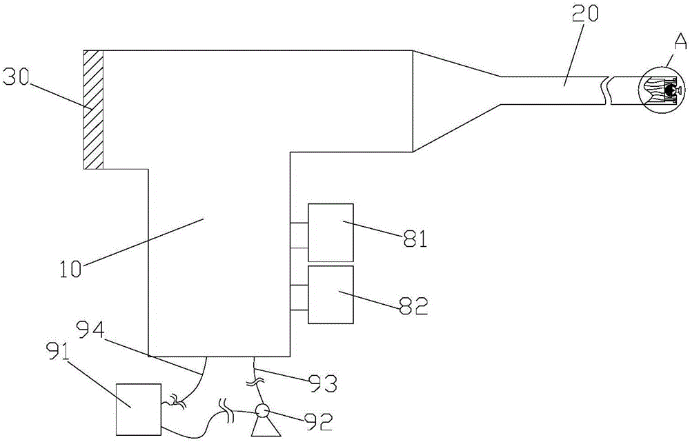
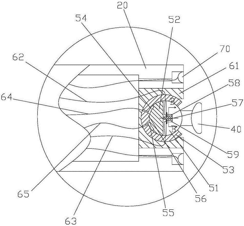
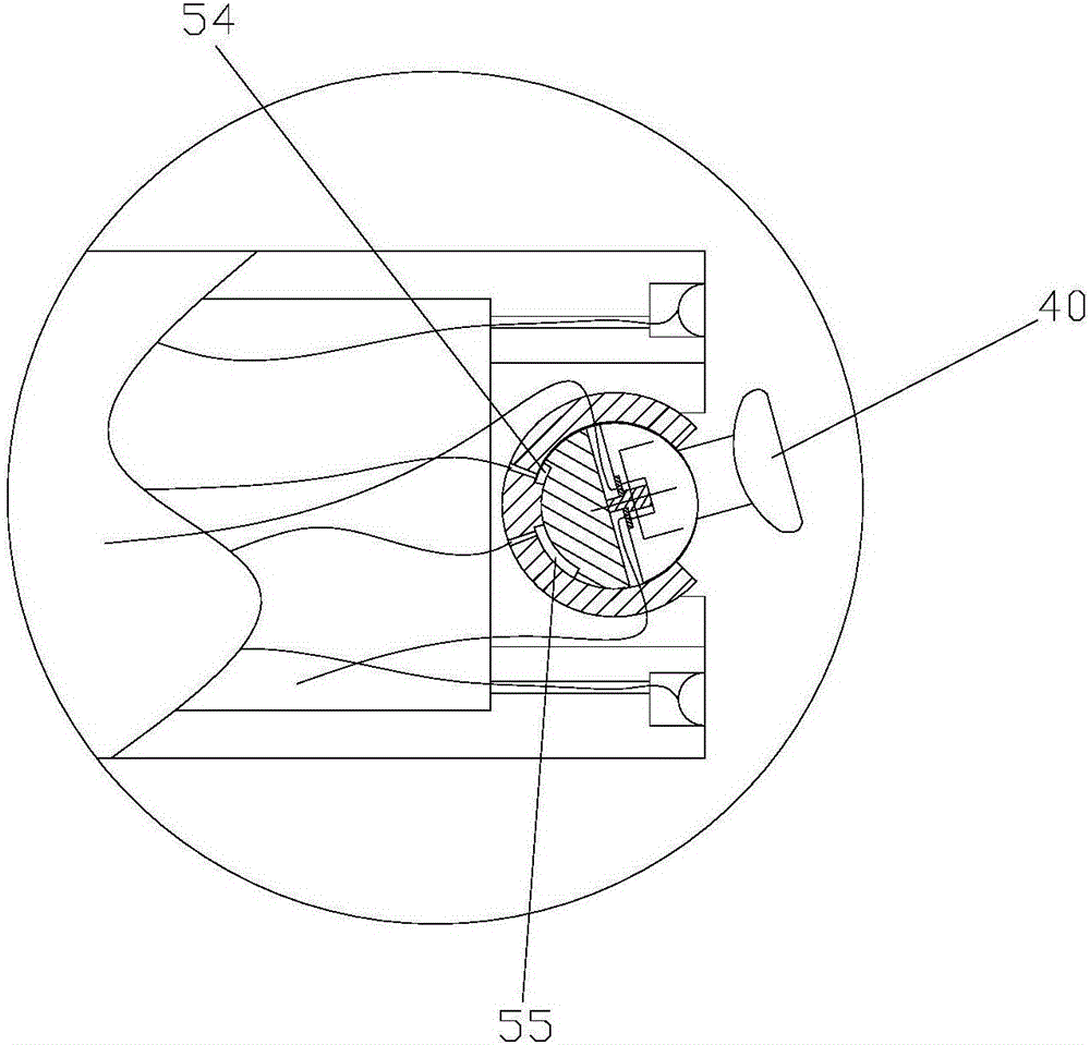
[0029] Such as Figures 1 to 5 As shown, a preferred embodiment of the present invention discloses an ENT inspection device for detecting the location of lesions in the ear canal, including: a main body 10, an intubation tube 20, an LED light 70, a ball bowl 53, a ball head, and a camera 40, display screen 30 and hydraulic drive mechanism. The intubation tube 20 is arranged at the front end of the main body 10 for insertion into the ear canal of the patient; the LED lamp 70 is arranged in the hole wall at the front end of the intubation tube 20 to provide a light source for exploration; the ball bowl 53 is arranged in the hole at the front end of the intubation tube 20 through the mount middle; the ball head is set in the ball b...


PUM


Abstract

The invention discloses an examination device for the otorhinolaryngological department, which is used in the position for detecting the focus in the auditory meatus. The device comprises a main body, an intubation tube arranged at the front end of the main body, an LED lamp arranged on the wall of the front end of the intubation tube, a ball fixed in the hole in the front end of the intubation tube, a bulb which is arranged in the ball and can rotate in the ball, a camera fixed on the bulb, a display screen which is arranged at the rear end of the main body and is used for displaying video images shot by the camera and a hydraulic drive mechanism which is used for driving the bulb to rotate, wherein the hydraulic drive mechanism is used for selectively introducing hydraulic medium to four cavities, so that the angle of the camera is adjusted under two degrees of rotational freedom, and the auditory meatus can be overall detected.

Description

technical field [0001] The invention relates to a medical device, in particular to an examination device for the department of otolaryngology. Background technique [0002] In the otolaryngology department of the hospital, the inspection instruments used to check the ear are very precise and light. The prior art provides a kind of otolaryngology inspection device for detecting whether there are lesions in the ear and the location of the lesions. The device includes a The main body, the intubation tube set at the front end of the main body for inserting into the ear canal, the camera set at the front end of the intubation tube for exploring the inside of the ear canal, and the display screen set at the rear end of the main body for displaying image information captured by the camera. Although the device can detect the internal situation of the ear canal through the camera, the camera cannot fully detect a certain position because the camera is fixed on the front end of the in...

Claims


Application Information

Patent Timeline
Patent Type & Authority Applications(China)
IPC IPC(8): A61B1/227A61B1/233A61B1/267A61B1/00A61B1/04
CPCA61B1/00045A61B1/04A61B1/0684A61B1/227A61B1/233A61B1/267
Inventor 厉媛媛
Owner 融创达科技成果转化(江苏)有限公司
Why Eureka
Try Eureka