Electronic Cystoscope

A technology of cystoscopy and electronics, which is applied in the field of electronic cystoscopy, can solve the problems of heavy insertion pain, long time-consuming diagnosis, and increased pain of patients, and achieve the effects of reducing production costs, good promotion significance, and improving accuracy

Active Publication Date: 2017-09-19
GUANGZHOU RED PINE MEDICAL INSTR CO LTD
View PDF8 Cites 3 Cited by
  • Summary
  • Abstract
  • Description
  • Claims
  • Application Information

AI Technical Summary

Problems solved by technology

The design of the existing cystoscope is not reasonable enough, the insertion is painful, the diagnosis takes a long time, and it also increases the pain suffered by the patient during the diagnosis process
Moreover, the manufacturing process of the image transmission fiber is complicated, resulting in high cost

Method used


Image

  • Electronic Cystoscope
  • Electronic Cystoscope
  • Electronic Cystoscope

Examples


Embodiment Construction

[0022] Embodiments of the present invention are described in detail below:

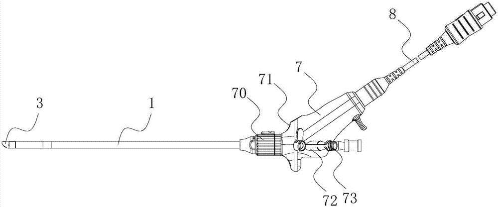
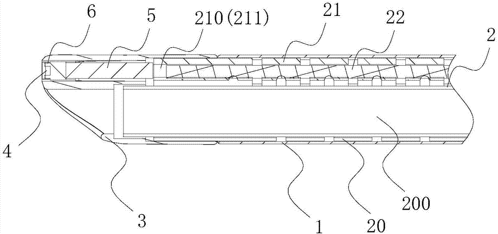
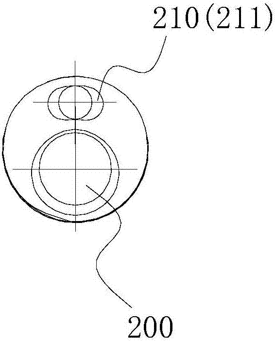
[0023] Such as Figure 1-3 As shown, the electronic cystoscope described in this embodiment includes a soft tube 1, a hard tube 2 and a display (not shown), the hard tube 2 is sleeved in the soft tube 1, and the hard tube 2 The tube 2 is provided with an instrument channel 200, an endoscopic channel 210 and an illumination channel 211. The endoscopic channel 210 is provided with a lens 4 and an image sensor 5. The illumination channel 211 is provided with a light source 6. The hard tube 2 is provided with an instrument hole communicating with the instrument channel 200, a lens hole communicating with the endoscopic channel 210, and a light exit hole communicating with the lighting channel 211. The lens hole matches the lens 4, and the light exit hole matches the light source 6. Matching, the image sensor 5 is electrically connected to the display.

[0024] The above-mentioned electronic cystoscope, ...


PUM


Abstract

The invention discloses an electronic cystoscope which comprises a soft tube, a hard tube and a displayer. The hard tube is sleeved with the soft tube, and an instrument channel, an endoscopy channel and an illumination channel are arranged in the hard tube; a lens and an image sensor are arranged in the endoscopy channel, and a light source is arranged in the illumination channel; the hard tube is provided with an instrument hole communicated with the instrument channel, a lens hole communicated with the endoscopy channel and a light-out hole communicated with the illumination channel; the lens hole is matched with the lens, the light-out hole is matched with the light source, and the image sensor is electrically connected with the displayer. According to the electronic cystoscope, by the adoption of the design that the hard tube is wrapped in the soft tube, the tube insertion softness is moderate, the pain of a patient is relieved, and the accuracy of a diagnosis result is improved. Meanwhile, the image sensor is used for replacing an existing image optical fiber, so that production cost is lowered, and the electronic cystoscope has extremely good popularization significance.

Description

technical field [0001] The invention relates to the field of medical instruments, in particular to an electronic cystoscope. Background technique [0002] Bladder examination is a common method used in urology to check urethral and bladder diseases, and it plays an important role in the diagnosis and treatment of urethral and bladder lesions. Cystoscope is an important tool for bladder examination and has been widely used clinically. When in use, the insertion tube of the cystoscope is inserted into the bladder along the curve of the urethra, and the image is transmitted to the eyepiece by means of the illumination fiber and the image transmission fiber. The design of the existing cystoscope is not reasonable enough, the insertion pain is great, the diagnosis takes a long time, and it also increases the pain suffered by the patient during the diagnosis process. Moreover, the manufacturing process of image transmission optical fiber is complicated, resulting in high cost. ...

Claims


Application Information

Patent Timeline
Patent Type & Authority Patents(China)
IPC IPC(8): A61B1/307A61B1/018A61B1/05A61B1/06A61B1/00
CPCA61B1/00135A61B1/018A61B1/05A61B1/0684A61B1/307
Inventor 叶章群
Owner GUANGZHOU RED PINE MEDICAL INSTR CO LTD
Why Eureka
Try Eureka