A Registration Method of Cardiac Perfusion Magnetic Resonance Images

A magnetic resonance imaging and heart technology, applied in the field of medical imaging, can solve problems such as low image contrast, limiting the reliability and efficiency of visual diagnosis

Active Publication Date: 2020-12-18
SHANGHAI UNITED IMAGING HEALTHCARE
View PDF3 Cites 0 Cited by
  • Summary
  • Abstract
  • Description
  • Claims
  • Application Information

AI Technical Summary

Problems solved by technology

However, deformation and motion artifacts in cardiac perfusion MR images caused by cardiac motion, respiration, and patient position movement during image acquisition, fast time sampling, and relatively low image contrast limit the potential for direct visual diagnosis. reliability and efficiency

Method used


Image

  • A Registration Method of Cardiac Perfusion Magnetic Resonance Images
  • A Registration Method of Cardiac Perfusion Magnetic Resonance Images
  • A Registration Method of Cardiac Perfusion Magnetic Resonance Images

Examples


Embodiment Construction

[0034] The present invention will be further described below in conjunction with the accompanying drawings and embodiments.

[0035] A registration method for cardiac perfusion magnetic resonance images, comprising the following steps:

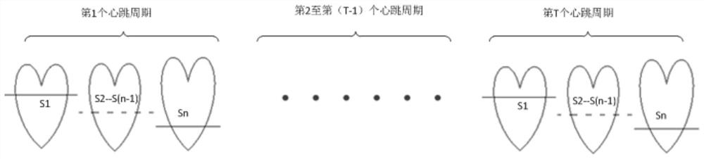
[0036] Acquisition of an MRI image containing several slices of the left ventricular myocardium I NT , where N represents the sequence number (number) of the layer where the slice is located in the same heartbeat cycle, T represents the sequence number (number) of different heartbeat cycles, and N and T are integers greater than or equal to 1; Magnetic resonance image of a slice of layer (M layer) I MT Carry out the following processing: the magnetic resonance image I of the selected starting layer (M=1) slice within T heartbeat cycles 1T , segmented MRI image I 1T In the endocardium, determine the magnetic resonance image I 1T The magnetic resonance image corresponding to the rth heartbeat cycle in is the reference image I 1r and the reg...


PUM


Abstract

The invention discloses a heart perfusion magnetic resonance image registration method. The method comprises steps: a magnetic resonance image I<NT> containing a plurality of layers of slices of left ventricular are acquired, a magnetic resonance image I<1T> for the initial layer of slice in T heartbeat cycles is selected, endocardium in the magnetic resonance image I<1T> is segmented, and a reference image I<1r> and a region of interest ROI<1> are determined; magnetic resonance images I<MT> for layers from the second to the Nth of slices in the T heartbeat cycles are selected respectively, a magnetic resonance image I<MT> for the current layer of slice the T heartbeat cycles is set, with the sequence number r of the heartbeat cycle of the reference image I<(M-1)r> of the former layer of slice and the region of interest ROI<M-1> as reference, the endocardium in the magnetic resonance image I<MT> for the Mth layer of slice is segmented, and a reference image I<Mr> in the magnetic resonance image I<MT> for the Mth layer of slice is acquired; and with the reference image for each layer of slice as reference, magnetic resonance image registration for the layer of slice in the T heartbeat cycles is completed.

Description

【Technical field】 [0001] The invention relates to the field of medical images, in particular to registration of cardiac perfusion magnetic resonance images. 【Background technique】 [0002] In recent years, the morbidity and mortality of cardiovascular diseases are increasing year by year. Tens of millions of people die of cardiovascular disease every year all over the world. Myocardial ischemia is the most important cause of cardiovascular morbidity and mortality. Cardiac perfusion magnetic resonance imaging is considered the first-line noninvasive test for ischemic heart disease. According to the basic principles of cardiac perfusion magnetic resonance images, radiologists can make a clinical diagnosis of myocardial ischemia by directly observing the changes in the signal intensity of the myocardial part of the left ventricle over time. However, deformation and motion artifacts in cardiac perfusion MR images caused by cardiac motion, respiration, and patient position mov...

Claims


Application Information

Patent Timeline
Patent Type & Authority Patents(China)
IPC IPC(8): G06T7/30G06T7/10
CPCG06T2207/10088G06T2207/20104
Inventor 姜娈凌姗李强
Owner SHANGHAI UNITED IMAGING HEALTHCARE
Why Eureka
Try Eureka